Spine Imaging
Mosby (Verlag)
978-0-323-03124-0 (ISBN)
- Titel erscheint in neuer Auflage
- Artikel merken
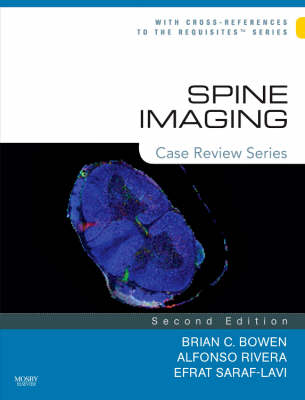
This volume in the best-selling "Case Review" series uses more than 200 case studies to challenge your knowledge on a full range of topics in spine imaging. Each case includes a set of 1 to 4 unknown images and four self-test questions, accompanied by answers, supporting literature references, and a commentary to help you gain a better understanding of how the correct diagnosis was reached. The discussion addresses the most important imaging, pathological, and clinical features of the case. This unique case-based format coupled with an easy-access organization and high-quality images equips you with the guidance you need to master the material, pass certification exams, and succeed in practice.
OPENING ROUND 1. Lumbar spinal stenosis 2. Diskitis-Osteo, lumbar 3. CSF flow imaging 4. Postoperative Recurrent disk herniation, lumbar 5. Unilateral facet dislocation (cervical) 6. Atlantoaxial rotatory deformity 7. Paget's disease 8. Traumatic vertebral artery occlusion 9. Caudal regression syndrome 10. Tarlov Cyst 11. Radial anular tear of a lumbar disk 12. Schwannoma, Sacral 13. Lateral meningocele, thoracic 14. Intradural lipoma, Cervical 15. Sagittal, epidural midline septum 16. Herniated disk constrained by midline septum 17. Loose transpedicular screw 18. Craniospinal tuberculosis 19. Disk herniation (extrusion), lumbar 20. Paget Disease 21. Sarcoidosis, cauda equina 22. Extradural schwannoma, cervical 23. Retroperitoneal sarcoma 24. Adult rheumatoid arthritis 25. Arachnoiditis 26. Spontaneous reduction of disk herniation 27. Intradural lipoma, conus. 28. Os odontoideum 29. Tuberculous Meningitis 30. Fracture-separation of the articular mass 31. Enhancing nerve roots with Lumbar disk herniation (unoperated spine) 32. Vertebral artery dissection 33. Lipoma of the Filum Terminale 34. Isthmic (Spondylolytic) spondylolisthesis 35. Baastrup's Disease 36. Multiple Meningiomas, Neurofibromatosis type 2 37. Sacral neurofibromas 38. Vertebroplasty 39. Atlantoaxial subluxation 40. Lipomyelomeningocele 41. Bilateral Transpedicular Ascending Lumbar Veins 42. Devic's syndrome (Neuromyelitis Optica) 43. Disk Herniation (Lateral) with Peridiskal Scar, Lumbar 44. Neuropathic Arthropathy (Charcot Spine) 45. Ossification of the posterior longitudinal ligament 46. Epidural abscess, thoracic 47. Epidural Steroid Injection 48. Odontoid nonunion 49. Disk Herniation, Cervical 50. Discogram demonstrating a left lateral (foraminal) disk herniation 51. Chiari II Malformation With Hydromyelia 52. Vertebral metastasis (prostate carcinoma) with epidural extension 53. Calcium pyrophosphate dihydrate (CPPD) crystal deposition 54. Intradural Meningioma, thoracic 55. Langerhans' Cell Histiocytosis with vertebra plana 56. Congenital arachnoid cyst (Type 1A meningeal cyst) 57. Leptomeningeal metastases (breast carcinoma) 58. Disk herniation (sequestration), lumbar 59. Ossification of the ligamentum flavum (OLF) 60. Diastematomyelia, two dural sacs 61. Epidural Lipomatosis 62. Aneurysmal Bone Cyst (C2) 63. Dural arteriovenous fistula 64. Sickle Cell Disease with osteomyelitis and epidural abscess (lumbar) 65. Chordoma, sacral 66. Multiple Sclerosis, cervical 67. Vertebral lymphoma with secondary epidural venous enlargement FAIR GAME 68. Intradural lipoma, lumbar 69. Traumatic Disk herniation, Cervical 70. Metastasis mimicking diskitis/osteomyelitis 71. CNS dissemination of conus ependymoma 72. Disk herniation (sequestration), cervical 73. Intradural, cystic teratoma 74. Fluid sign 75. Chronic Inflammatory Demyelinating Polyradiculoneuropathy (CIDP) 76. Steroid-Induced, Chronic Osteoporotic Compression Fractures 77. Pediatric spine, normal MR signal intensities 78. multiple sclerosis mimicking tumor 79. Bilateral facet dislocation with central cord syndrome 80. Dislodged bone graft (posterior) 81. Arteriovenous Malformation (Juvenile Type at C1-C2) 82. Myxopapillary ependymoma 83. Hemangioblastoma, Upper Thoracic 84. Benign vertebral hemangioma 85. Terminal Ventricle and Syringohydromyelia 86. Sacrococcgeal teratoma 87. Osteoid osteoma, lumbar 88. Intradural meningioma, thoracic 89. lymphoma 90. Iophendylate (Pantopaque) Collection, thoracic 91. Arachnoid cyst (postsurgical), thoracic 92. Guillain-Barre syndrome 93. Chordoma, cervical 94. Sarcoidosis, cervical 95. Osteoblastoma, cervical 96. Enterogenous cyst 97. Muscular dystrophy 98. Sacral Insufficiency Fractures and Sacroplasty 99. Lumbar juxtaarticular (synovial) cyst 100. Ependymoma, Cervical 101. Malignant peripheral nerve sheath tumor 102. Cavernous Malformation 103. Primary Vertebral and Epidural Lymphoma 104. Leukemia 105. Spinal Cord Infarction, Thoracic 106. Neurofibomatosis type 1, with cord compression 107. Intradural schwanomma, thoracic 108. Disk Herniation, Thoracic 109. Intramedullary metastases (breast carcinoma) 110. Cryptococcosis 111. Ankylosing spondylitis with dural ectasia and arachnoiditis 112. Traumatic pseudomeningocele 113. Atlanto-occipital Assimilation with Basilar Invagination 114. Acute multiple sclerosis 115. Amyotrophic Lateral Sclerosis 116. Hemangioblastoma of the filum terminale 117. Chiari I malformation 118. Degenerative Facet Synovitis, Cervical 119. Glioblastoma multiforme seeding the spinal subarachnoid space 120. Ependymoma with cyst, cervical 121. Intramedullary Arteriovenous Malformation (Glomus Type at T12) 122. Cranial Settling 123. Epidermoid, Lumbar 124. Epidural Abscess, Thoracolumbar 125. Metastatic Tumor Infiltration of Sacral Nerves and Plexus 126. Conus Medullaris Infarction 127. Dural Arteriovenous Fistula (T11) CHALLENGE 128. Intradural myolipoma, thoracic 129. Wallerian degeneration, cervical 130. Vertebral body avascular necrosis 131. Unilateral Cord Infarction, Cervical 132. Acute spinal subdural hematoma 133. Intraspinal Gas Collection 134. Radiation myelopathy 135. Polyostotic Fibrous Dysplasia 136. Subacute combined degeneration (SCD) 137. Listeria myelitis/rhombencephalitis 138. Retrosomatic cleft 139. Acute calcific prevertebral tendenitis (calcific tendenitis of the longus colli muscle) 140. Charcot-Marie-Tooth Disease (Hereditary Motor and Sensory Neuropathy [Type I]) 141. Expansive open-door laminaplasty 142. Plasmacytoma 143. Vestigial Tail 144. Retroisthmic cleft 145. Atlantoaxial dislocation with occipitalized atlas 146. Intramedullary abscess 147. Laryngeal Tuberculosis and Tuberculous Spondylitis 148. Acute vertebral body infarcts 149. Subarachnoid Hemorrhage With Organized Hematoma 150. Adult polyglucosan body disease 151. Varicella-Zoster Virus Myelitis 152. Progressive Spinal Muscular Atrophy 153. Dejerine-Sottas Disease (Hereditary Motor and Sensory Neuropathy Type III) 154. Schmorl's node hiding a metastasis 155. Spontaneous, acute epidural hematoma 156. Scheuermann Kyphosis 157. Primary CNS lymphoma 158. Aggressive vertebral hemangioma 159. Epidural angiolipoma 160. Kyphoplasty with associated diskitis/osteomyelitis 161. Acute Disseminated Encephalomyelitis 162. Hematomyelia (acute) secondary to an ependymoma 163. Neuroepithelial cyst 164. Ganglioneuroma 165. Lumbosacral trunk enlargement 166. Intradural Schwannoma, Cervical 167. Calcified cervical disc herniation in a child 168. Ossifying Fibroma 169. Ruptured perineurial cyst, thoracic 170. Pantopaque Mimicking Intradural Lipoma 171. Subacute combined degeneration (SCD). 172. Recurrent Lumbar Dermoid 173. Seronegative Spondyloarthritis (Crohn Disease) 174. Radiation-Induced Malignant Fibrous Histiocytoma 175. Paraganglioma of the Cauda Equina 176. Intraspinal Extradural Arachnoid Cyst (Lymphedema-Distichiasis Syndrome) 177. CSF hypotension (and hypovolemia) 178. Chordoma, thoracic 179. Meningeal Melanocytoma 180. Congenital Absence of the Pedicle 181. Subdural empyema 182. Diastematomyelia, Two Dural Sacs 183. Spinal hydatid cyst
| Reihe/Serie | Case Review Series |
|---|---|
| Zusatzinfo | Approx. 420 illustrations |
| Verlagsort | St Louis |
| Sprache | englisch |
| Maße | 211 x 276 mm |
| Themenwelt | Medizinische Fachgebiete ► Chirurgie ► Neurochirurgie |
| Medizin / Pharmazie ► Medizinische Fachgebiete ► Neurologie | |
| Medizin / Pharmazie ► Medizinische Fachgebiete ► Radiologie / Bildgebende Verfahren | |
| Schlagworte | Bildgebende Verfahren (Medizin) • Wirbelsäule |
| ISBN-10 | 0-323-03124-2 / 0323031242 |
| ISBN-13 | 978-0-323-03124-0 / 9780323031240 |
| Zustand | Neuware |
| Haben Sie eine Frage zum Produkt? |
aus dem Bereich


