Spine Imaging
Mosby (Verlag)
978-0-323-00507-4 (ISBN)
- Titel erscheint in neuer Auflage
- Artikel merken
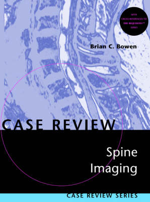
This new volume in the popular Case Review series presents 175 cases of spinal pathology and anatomic variants. The clinical images represent all diagnostic modalities, including MR, CT, plain radiographs, angiograms, and myelograms. Each case begins with a set of images and four self-test questions, followed by the answers, supporting literature references, and a comment. The comment addresses the most important imaging, pathological, and clinical features of the case.
Brian C. Bowen, M.D., Ph.D., is Associate Professor of Radiology and Neurological Surgery at the University of Miami School of Medicine and Attending Neuroradiologist at the Jackson Memorial Medical Center. He is an up-and-coming star in neuroradiology and is affiliated with a radiology department chaired by the prestigious Dr. Robert Quencer. He is extremely active with regard to publishing journal articles, book chapters, and with regard to presenting exhibits and lectures. As a tribute to his reputation and expertise in spine imaging, Drs. David Stark and Bill Bradley asked him to contribute two chapters to the spine section of the third edition of Magnetic Resonance Imaging. Dr. Bowen is the holder of numerous awards, some from the National Institute of Health and the National Science Foundation. He is an active teacher, lecturer,thesis advisor, and member of the Task Force for Education at his school.
Dural AVM Intramedullary AVM Cavernous angioma Arachnoid cyst (post-infectious), thoracic Epidural lipomatosis Diastematomyelia, single dural sac Diastematomyelia, two dural sacs Epidural abscess Epidermoid cyst Disk herniation (sequestration), lumbar Conus medullaris infarction Thoracic cord infarction Leptomeningeal metastases (melanoma) Hemangioblastoma Degenerative facet disease, cervical Congenital absence of the pedicle Chiari I malformation, pre- and post-treatment Diffuse hypertrophic skeletal hyperostosis (DISH), with fracture Langerhans cell histocytosis Amyotrophic lateral sclerosis Subacute necrotizing myelopathy Acute multiple sclerosis, cervical cord Multilevel osteomyelitis/diskitis Cervical astrocytoma Intramedullary metastasis (melanoma) Intradural meningioma, thoracic Drop metastases (glioblastoma multiforme) Traumatic pseudomeningocele Vertebral metastasis (colon carcinoma) with epidural extension Chordoma, thoracic Ankylosing spondylitis with dural ectasia Intramedullary metastasis (breast carcinoma) Chronic coccidioidal meningitis with syringomyelia Chiari II malformation with hydromyelia Disk herniation, thoracic Disk herniation, cervical Odontoid fracture, Type II Os odontoideum Anterior sacral meningocele CSF pulsation artifacts Intradural schwannoma, thoracic Ossification of the posterior longitudinal ligament Thoracic ependymoma Inhomogeneous fat suppression: sacral met NF1 with cord compression Cervical ependymoma with syringohydromyelic cavity Lumbar herniated disk, lateral Devic's syndrome Lipomyelomeningocele Basilar invagination Intramedullary AVM (T1 level) Filum ependymoma Odontoid chordoma Primary vertebral and epidural lymphoma Neuroblastoma Adult tethered cord with a dermal sinus Intraspinal extradural arachnoid cyst (lymphedema-distichiasis syndrome) Atlanto-axial subluxation Radiation-induced malignant fibrous histiocytoma Sacral neurofibromas (neurofibromatosis type 1) Metastatic tumor infiltration of sacral nerves and plexus Malignant peripheral nerve sheath tumor Perineural spread of rectosigmoid carcinoma along sacral nerves Multiple spinal meningiomas Lumbar synovial cyst Isthmic (spondylolytic) spondylolisthesis Hodgkin's lymphoma, post-chemotherapy Dermal sinus tract Acute myeloid leukemia with granulocytic sarcoma (chloroma) Diskitis/osteomyelitis, pediatric Lipoma of the filum terminale Enhancing nerve roots with lumbar disk herniation (unoperated spine) Fatty replacement in lumbar paraspinous muscles Fracture separation of the articular mass with unilateral "perched" facet Extradural meningioma, thoracic Enterogenous cyst Osteoblastoma, cervical Juvenile rheumatoid arthritis Recurrent lumbar dermoid Epidural abscess, thoracic Pantopaque mimicking intradural lipoma Hemangioendothelioma, thoracic Sarcoidosis, cervical Ossifying fibroma, lumbar Intradural schwannoma, cervical Intradural lipoma, thoracic Burst fracture with acute epidural hematoma, lumbar Chordoma, cervical Arachnoid cyst (post-surgical), thoracic Arachnoiditis Platybasia with syringohydromyelia Iophendylate (Pantopaque) collection Ganglioneuroma, lumbosacral Vascular malformation Phased-array coil (multicoil) system Adult rheumatoid arthritis Syringohydromyelia, postinflammatory Anterior sacral meningocele Lymphoma Extradural schwannoma, cervical Systemic lupus erythematosis, with intramedullary hemorrhage Neuroepithelial cyst, cervical Sarcoidosis, cauda equina Acute disseminated encephalomyelitis (ADEM), cervicothoracic Meningioma, cervical Herniated disk with cephalad migration, lumbar Leptomeningial carcinomatosis Osteoid osteoma, lumbar Epidural angiolipoma Craniospinal tuberculosis Aggressive vertebral hemangioma Multiple meningiomas, thoracic Magnetic susceptibility effects (blooming artifact) Intramedullary lymphoma Lateral meningocele, thoracic Terminal ventricle/terminal syringohydromyelia Spontaneous, acute epidural hematoma, thoracolumbar Post-operative intramedullary cyst with flow Multiple vertebral hemangiomas Progressive post-traumatic myelomalacic myelopathy, cervical Vacuolar myelopathy, thoracic Hamartoma of the conus medullaris (neurofibromatosis, type 1) Herpes zoster myelitis Toxoplasmic myelitis Intraspinal bullet and post-traumatic arachnoiditis Metastatic melanoma, epidural and intradural Subarachnoid hemorrhage with organized hematoma Leptomeningeal metastases simulating intramedullary tumor Epidural abscess/cellulitis, cervical Laryngeal tuberculosis with tuberculous spondylitis Spinal cord AVM (juvenile), cervical Paget's disease, lumbar Bilateral facet dislocation with central cord syndrome, cervical Traumatic disk herniation, cervical Intramedullary abscess, conus medullaris Intramedullary mass (ependymoma) Dejerine-Sottas disease, cauda equina Sagittal, epidural midline septum, lumbar Herniated disk constrained by midline septum Subdural injection of contrast Charcot-Marie-Tooth disease, cauda equina Pediatric spine, normal MR signal intensities Steroid-induced, chronic osteoporotic compression fractures Schwannoma, sacral Plasmacytoma, cervical Degenerative changes in vertebral body marrow ("end-plate changes") Chronic inflammatory demyelinating polyradiculoneuropathy (CIDP) Expansive open-door laminaplasty, cervical Omental myelosynangiosis, cervical Dermoid CT myelography Retroisthmic cleft Retrosomatic cleft Accessory ossicle, facet Tarlov cysts Post-operative cord tethering, cervical Caudal regression syndrome Cervical HNP, free fragment Vertebroplasty Rotatory subluxation B12 deficiency Polyostotic fibrous dysplasia Metastatic ependymoma Metastasis Multiple sclerosis mimicking tumor Gas collection in a herniated disk Cord infarction, cervical Epidural metastasis Multiple sclerosis Postoperative, recurrent disk herniation CSF flow imaging Chordoma, sacral Wallerian degeneration Sickle cell disease with infection Benign vertebral fracture
| Erscheint lt. Verlag | 21.9.2001 |
|---|---|
| Reihe/Serie | Case Review Series |
| Zusatzinfo | 350 ills |
| Verlagsort | St Louis |
| Sprache | englisch |
| Maße | 216 x 279 mm |
| Gewicht | 755 g |
| Themenwelt | Medizinische Fachgebiete ► Chirurgie ► Unfallchirurgie / Orthopädie |
| Medizin / Pharmazie ► Medizinische Fachgebiete ► Radiologie / Bildgebende Verfahren | |
| ISBN-10 | 0-323-00507-1 / 0323005071 |
| ISBN-13 | 978-0-323-00507-4 / 9780323005074 |
| Zustand | Neuware |
| Haben Sie eine Frage zum Produkt? |
aus dem Bereich


