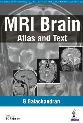
MRI Brain
Atlas and Text
Seiten
2015
Jaypee Brothers Medical Publishers (Verlag)
978-93-5250-022-2 (ISBN)
Jaypee Brothers Medical Publishers (Verlag)
978-93-5250-022-2 (ISBN)
- Titel nicht im Sortiment
- Artikel merken
Covers a wide spectrum of neuroradiology cases seen daily practice. As well as images and line diagrams, there is a brief interpretation of images, description of the case findings and relevant references for further reading. Useful tables are also included.
MRI Brain: Atlas and Text is a highly illustrated collection of magnetic resonance imaging cases, complete with guidance on terminology, anatomy and diagnosis.
Divided into five sections, the book begins with the basics of MRI, followed by an illustrated chapter on normal cross sectional MRI anatomy of the brain, MRI signals and sequences, and tumour diagnosis using MRI. The book concludes with an atlas of MRI cases, with 413 high quality MR images of the brain across 100 cases.
Each evidence based neuroradiology case begins with high quality MR images followed by discussion on the case findings, and concluded by relevant references for further reading.
MRI Brain: Atlas and Text covers MR signal intensity nomenclature, common MR sequences and their use, and the use of MRI in the diagnosis of stroke, along with other specialist topics making this book ideal for radiology postgraduates as well as GPs and neuroradiologists.
Key Points
Highly illustrated guide to magnetic resonance imaging
Features 100 evidence based MRI cases with high quality images, case findings and further reading
428 full colour images and illustrations
G Balachandran MD, DNB, DMRD, FICR Associate Professor of Radiology, Sri Manakula Vinayagar Medical College and Hospital, Pondicherry
MRI Basics: Physics
Normal Cross Sectional MR Anatomy of Brain
MRI of Signals and Sequences
Principles of Intracranial Space Occupying Lesions (ICSOL)/Tumor Diagnosis by MRI
MRI Atlas of Cases
| Erscheinungsdatum | 02.12.2015 |
|---|---|
| Verlagsort | New Delhi |
| Sprache | englisch |
| Maße | 159 x 241 mm |
| Gewicht | 450 g |
| Themenwelt | Medizin / Pharmazie ► Medizinische Fachgebiete ► Neurologie |
| Medizinische Fachgebiete ► Radiologie / Bildgebende Verfahren ► Kernspintomographie (MRT) | |
| ISBN-10 | 93-5250-022-9 / 9352500229 |
| ISBN-13 | 978-93-5250-022-2 / 9789352500222 |
| Zustand | Neuware |
| Haben Sie eine Frage zum Produkt? |
Mehr entdecken
aus dem Bereich
aus dem Bereich
Lehrbuch und Fallsammlung zur MRT des Bewegungsapparates
Buch | Hardcover (2020)
mr-verlag
CHF 306,55


