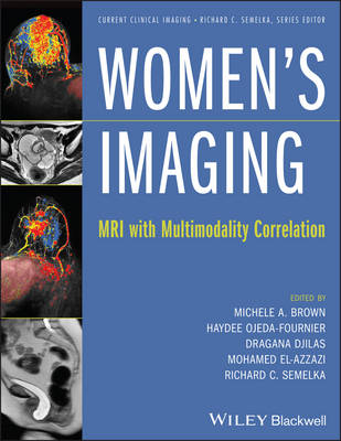
Women's Imaging
Wiley-Blackwell (Verlag)
978-1-118-48284-1 (ISBN)
The first complete reference dedicated to the full spectrum of women's imaging topics
"Women’s imaging" refers to the use of imaging modalities (X-ray, ultrasound, CT scan, and MRI) available for aiding in the diagnosis and care of such female-centric diseases as cancer of the breast, uterus, and ovaries. Currently, there is no single reference source that provides adequate discussions of MRI and its important role in the diagnosis of patients with women's health issues.
Thoroughly illustrated with the highest-quality radiographic images available, Women’s Imaging: MRI with Multimodality Correlation provides a concise overview of the topic and emphasizes practical image interpretation. It makes clear use of tables and diagrams, and offers careful examination of differential diagnosis with special notes on key learning points. Placing great emphasis on magnetic resonance imaging (MRI), while providing correlations to other important imaging modalities, the comprehensive book features the latest guidelines on imaging screening and includes in-depth chapter coverage of:
Pelvis MRI: Introduction and Technique
Imaging the Vagina and Urethra
Pelvic Floor Imaging
Imaging the Uterus
Imaging the Adnexa
Imaging Maternal Conditions in Pregnancy
Fetal Imaging
Breast MRI: Introduction and Technique
ACR Breast MRI Lexicon and Interpretation
Preoperative Breast Cancer Evaluation and Advanced Breast Cancer Imaging
Postsurgical Breast and Implant Imaging
MR-Guided Breast Interventions
Providing up-to-date information on many of the health issues that affect women across the globe, Women's Imaging will appeal to all general radiologists – especially those specializing in body imaging, breast imaging, and women’s imaging – as well as gynaecologists and obstetricians, breast surgeons, oncologists, radiation oncologists, and MRI technologists.
Mich?le Brown, MD, is Associate Professor of Radiology at UCSD Medical Center in San Diego, California, where she has practiced clinical radiology specializing in female pelvis imaging for over 10 years. She has written many articles and book chapters on various topics in the field of women's imaging, and she coauthored a well-received book on the subject of pelvic MRI. This is her first book as senior editor. Richard Semelka, MD, is an eminent senior radiologist and widely published book and journal article author and editor. A professor of radiology at the University of North Carolina at Chapel Hill, he is well known to Wiley-Blackwell customers as the editor for the best-selling monumental 2-volume work, Abdominal-Pelvic MRI, now in its third edition. He is also Series Editor for the book series for which the present new volume is intended. Dragana Djilas-Ivanovic, MD, is a professor and clinical radiologist at the Center forDiagnostic Imaging, Oncology Institute of Vojvodina in Novi Sad, Serbia. Having trained in the United States and various European nations in addition to Serbia, she is the author of over 70 original research articles and numerous book chapters dealing with women's imaging and soft tissue tumors. Mohamed Elazzazi, MD, PhD, is a professor and clinical radiologist in the Al-Azhar University in Cairo, Egypt, and an Associate Professor in the Department of Radiology, University of North Carolina at Chapel Hill.
Contributors, vii
Preface, ix
1 Pelvis MRI: introduction and technique, 1
Michele A. Brown & Richard C. Semelka
2 Imaging the vagina and urethra, 8
Shannon St. Clair, Randy Fanous, Mohamed El-Azzazi, Richard C. Semelka, & Michele A. Brown
3 Pelvic floor imaging, 27
Laura E. Rueff & Steven S. Raman
4 Imaging the uterus, 49
Randy Fanous, Katherine M. Richman, Chayanin Angthong, Mohamed El-Azzazi, & Michele A. Brown
5 Imaging the adnexa, 88
Michele A. Brown, Mary K. O’Boyle, Chayanin Angthong, Mohamed El-Azzazi, & Richard C. Semelka
6 Imaging maternal conditions in pregnancy, 131
Lorene E. Romine, Randy Fanous, Michael J. Gabe, Richard C. Semelka, & Michele A. Brown
7 Fetal imaging, 180
Lorene E. Romine, Ryan C. Rockhill, Michael J. Gabe, Reena Malhotra, Richard C. Semelka, & Michele A. Brown
8 Breast MRI: introduction and technique, 239
Michael J. Gabe, Jasmina Boban, Dragana Djilas, Vladimir Ivanovic, & Haydee Ojeda-Fournier
9 ACR breast MRI lexicon and interpretation, 264
Julie Bykowski, Natasa Prvulovic Bunovic, Dragana Djilas, & Haydee Ojeda-Fournier
10 Preoperative breast cancer evaluation and advanced breast cancer imaging, 296
Jade de Guzman, Dragana Bogdanovic-Stojanovic, Dragana Djilas, & Haydee Ojeda-Fournier
11 Postsurgical breast and implant imaging, 322
Julie Bykowski, Dag Pavic, Dragana Djilas, & Haydee Ojeda-Fournier
12 MR-guided breast interventions, 346
Michael J. Gabe, Dragana Djilas, Dag Pavic, & Haydee Ojeda-Fournier
Index, 363
| Reihe/Serie | Current Clinical Imaging |
|---|---|
| Verlagsort | Hoboken |
| Sprache | englisch |
| Maße | 224 x 287 mm |
| Gewicht | 1311 g |
| Themenwelt | Medizin / Pharmazie ► Medizinische Fachgebiete ► Gynäkologie / Geburtshilfe |
| Medizinische Fachgebiete ► Radiologie / Bildgebende Verfahren ► Kernspintomographie (MRT) | |
| ISBN-10 | 1-118-48284-0 / 1118482840 |
| ISBN-13 | 978-1-118-48284-1 / 9781118482841 |
| Zustand | Neuware |
| Informationen gemäß Produktsicherheitsverordnung (GPSR) | |
| Haben Sie eine Frage zum Produkt? |
aus dem Bereich


