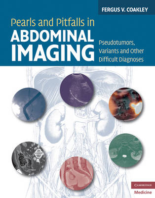
Pearls and Pitfalls in Abdominal Imaging
Pseudotumors, Variants and Other Difficult Diagnoses
Seiten
2010
Cambridge University Press (Verlag)
978-0-521-51377-7 (ISBN)
Cambridge University Press (Verlag)
978-0-521-51377-7 (ISBN)
Presents over 100 conditions, illustrated with over 700 figures, in the abdomen and pelvis which can commonly cause confusion and mismanagement in daily radiological practice, providing a focused textbook that can be readily used to avoid wrong diagnoses and prevent incorrect management or even malpractice litigation.
Research consistently suggests that 1.0 to 2.6% of radiology reports contain serious errors, many of which are avoidable, and it is clear that all radiologists can struggle with the basic questions as to whether a study is normal or abnormal. Pearls and Pitfalls in Abdominal Imaging presents over 100 conditions in the abdomen and pelvis which can commonly cause confusion and mismanagement in daily radiological practice, providing a focused textbook that can be readily used to avoid wrong diagnoses and prevent incorrect management or even malpractice litigation. It includes 700 figures and covers all the major modalities including CT, PET/CT and MRI. The pearls and pitfalls include artifacts, anatomic variants, mimics, and a miscellany of diagnoses that are under-recognized or only recently described. Conditions are presented in anatomic order from diaphragm to the symphysis pubis, with grouping by location and organ system.
Research consistently suggests that 1.0 to 2.6% of radiology reports contain serious errors, many of which are avoidable, and it is clear that all radiologists can struggle with the basic questions as to whether a study is normal or abnormal. Pearls and Pitfalls in Abdominal Imaging presents over 100 conditions in the abdomen and pelvis which can commonly cause confusion and mismanagement in daily radiological practice, providing a focused textbook that can be readily used to avoid wrong diagnoses and prevent incorrect management or even malpractice litigation. It includes 700 figures and covers all the major modalities including CT, PET/CT and MRI. The pearls and pitfalls include artifacts, anatomic variants, mimics, and a miscellany of diagnoses that are under-recognized or only recently described. Conditions are presented in anatomic order from diaphragm to the symphysis pubis, with grouping by location and organ system.
Fergus V. Coakley is Professor of Radiology and Urology, Section Chief of Abdominal Imaging, and Vice Chair for Clinical Services, Department of Radiology and Biomedical Imaging, University of California, San Francisco, CA, USA.
Preface; Diaphragm and adjacent structures; Liver; Biliary system; Spleen; Pancreas; Adrenal glands; Kidneys; Retroperitoneum; Gastro-intestinal tract; Peritoneal cavity; Ovaries; Uterus and vagina; Bladder; Pelvic soft tissues; Groin; Bones; Index.
| Erscheint lt. Verlag | 14.10.2010 |
|---|---|
| Zusatzinfo | 28 Halftones, color; 714 Halftones, black and white |
| Verlagsort | Cambridge |
| Sprache | englisch |
| Maße | 225 x 282 mm |
| Gewicht | 1530 g |
| Themenwelt | Medizinische Fachgebiete ► Radiologie / Bildgebende Verfahren ► Kernspintomographie (MRT) |
| Medizinische Fachgebiete ► Radiologie / Bildgebende Verfahren ► Radiologie | |
| ISBN-10 | 0-521-51377-4 / 0521513774 |
| ISBN-13 | 978-0-521-51377-7 / 9780521513777 |
| Zustand | Neuware |
| Haben Sie eine Frage zum Produkt? |
Mehr entdecken
aus dem Bereich
aus dem Bereich
Lehrbuch und Fallsammlung zur MRT des Bewegungsapparates
Buch | Hardcover (2020)
mr-verlag
CHF 306,55


