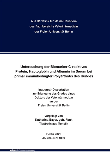
Untersuchung der Biomarker C-reaktives Protein, Haptoglobin und Albumin im Serum bei primär immunbedingter Polyarthritis des Hundes
Seiten
2023
|
1. Auflage
Mensch & Buch (Verlag)
978-3-96729-188-9 (ISBN)
Mensch & Buch (Verlag)
978-3-96729-188-9 (ISBN)
- Keine Verlagsinformationen verfügbar
- Artikel merken
Bei der primär immunbedingten Polyarthritis (IPA-I) des Hundes handelt es sich um eine entzündliche Gelenkerkrankung nicht-infektiöser Ätiologie, gekennzeichnet durch eine Synovitis mit oftmals systemischen Krankheitszeichen wie Lethargie, Arthralgie und Fieber.
Akute Phase Proteine werden zur Diagnose, Prognose und zur Überwachung des Therapieerfolges bei verschiedensten Erkrankungen eingesetzt. Zur Verwendung als Biomarker bei IPA-I des Hundes gibt es bisher nur wenige Daten.
Das Ziel dieser prospektiven Studie war es, das C-reaktive Protein (CRP), Haptoglobin und Albumin im Verlauf bei Hunden mit IPA-I zu messen, mit dem klinischen Verlauf unter Therapie zu vergleichen und ihren Nutzen bezüglich Diagnostik, Monitoring und Prognose zu evaluieren. Dazu wurden von Oktober 2014 bis Dezember 2019 an der Klinik für kleine Haustiere der Freien Universität Berlin 21 Hunde mit IPA-I, bei denen eine vollständige diagnostische Abklärung inkl. Gelenkpunktion, Synoviaanalyse, Blutbild, Blutchemie, Tests auf Infektionserkrankungen, Röntgen, Sonografie und regelmäßige Verlaufskontrollen möglich waren, eingeschlossen. Bei jedem Kontrolltermin wurden klinische und Laborkontrollen durchgeführt und die CRP-, Haptoglobin-, und Albumin- Konzentrationen im Serum gemessen. Alle Patienten wurden bei Erstvorstellung (T0) beprobt. Weitere Seren wurden bei jeweils 20/21 Hunden (95,2 %) an Tag 2-7 (T1) und Tag 8-14 (T2) gesammelt. Bei 19/21 Hunden (90,5 %) wurden weitere Kontrollen in den Wochen 3–4 (T3) und bei 16/21 (76,2 %) Hunden in Woche 5-8 (T4) durchgeführt. Bei 12/21Tieren (57,1 %) erfolgten Kontrollen über die genannten Zeitpunkte hinaus.
Die erhobenen Daten wurden mittels Shapiro-Wilk-Test auf Normalverteilung überprüft und deskriptiv ausgewertet. Bei nicht normalverteilten Daten wurden nicht-parametrische Tests (Wilcoxon-Test, Friedman-Test), bei normalverteilten Daten parametrische Tests (T-Test, ANOVA) angewendet. Das Signifikanzniveau wurde auf α ≤0,05 festgesetzt.
Die klinischen Symptome (Dauer 1-300 Tage) bei Erstvorstellung umfassten Fieber (20/21; 95,2 %), gestörtes Allgemeinbefinden (19/20; 90,5 %), Lahmheit (14/21; 66,6 %), vermehrte Gelenkfüllung (12/21; 57,1 %), Gelenkbiegeschmerz (13/21; 61,9 %), vermehrt erwärmte Gelenke (10/21; 47,6 %), Halsbiegeschmerz/Dolenz der Wirbelsäule (4/21; 19 %).
Die Röntgenaufnahmen ergaben bei 13/21 Hunden (61,9 %) einen vermehrten Kapsel-/Weichteilschatten an mindestens einem Gelenk.
Zu den entzündlich veränderten Gelenken gehörten: Karpus (28/31 punktierten Karpalgelenken; 90,3 %), Knie (35/39, 89,7 %), Tarsus (24/30; 80 %), Ellenbogen (18/23; 78,2 %), Schulter (2/2).
Nach Diagnosestellung erfolgte eine immunsuppressive Therapie mit Prednisolon bei allen Hunden. Zusätzlich erhielten zehn Hunde Leflunomid, drei Ciclosporin und einer Mycophenolat-mofetil. Es besserten sich daraufhin alle Hunde klinisch.
Alle Hunde wurden an T0, 20/21 (95,2 %) an T1 und T2, 19/21 (90,5 %) an T3 und 16/21 (76,2 %) an T4 nachuntersucht. Bei allen Hunden war CRP an T0 erhöht (16–169 mg/l, median [m] 97,1; Leukozytenzahl [WBC] 6,7-64,7 G/l [m 15,5]; Albumin [Alb] 26,1 g/l [SD±3,4]). Im Verlauf fiel bei allen Hunden die CRP-Konzentration von T0 zu T4 ab (p<0,0001), bei der Leukozytenzahl ergab sich keine signifikante Konzentrationsänderung, während Albumin zu Beginn bei 13/21 (62 %) erniedrigt war und im Verlauf anstieg (T1: CRP 3,5-149,9 mg/l [m 26,6]; WBC 9,1-62,2 G/l [m 20,8]; Alb 28,2 g/l [SD±3,3 g/l]; T2: CRP 0,3-88,6 mg/l [m 7,4]; WBC 6,7-73,4 G/l [m 19,3]; Alb 29,3 g/l [SD±2,6]); T3: CRP 0,2-100,1 mg/l [m 3,0]; WBC 8,1-53,9 G/l [m 17,0], Alb 30,9 g/l [SD±2,4]). An T4 lag CRP bei 19/21 Hunden im Referenzbereich (0,5-38,5 mg/l [m 1,8]; WBC 7,0-37,5 G/l [m 12,8]; Alb 30,7 g/l [SD±1,8 g/l]). Bei 8/21 Hunden (38,1 %) kam es zum Rezidiv. Das CRP stieg signifikant an (19,9-159,4 mg/l [m 66,3]) und fiel nach erfolgreicher Therapie erneut ab. Die Konzentrationsänderungen der Leukozytenzahl, Albumin waren nicht signifikant.
Keiner der Hunde zeigte einen erhöhten Haptoglobin-Wert an T0. Die Differenzen des Haptoglobins zwischen T0 zu T4 waren statistisch nicht signifikant (p= 0,93) und auch im Verlauf konnte kein signifikanter Unterschied des Haptoglobins an den verschiedenen Zeitpunkten festgestellt werden.
Limitationen der Studie sind die relativ kleine Studienpopulation, die unterschiedlichen Zeitpunkte der Probenentnahme und die Anzahl der Proben je Tier.
Basierend auf den Daten der vorliegenden klinischen Studie wurde deutlich, dass CRP bei an IPA-I erkrankten Hunden als Biomarker hinsichtlich Diagnostik und Monitoring Verwendung finden kann. Der schnelle Konzentrationsanstieg des CRPs während der Verlaufsmessungen spiegelt die ablaufende Akute-Phase-Reaktion und somit die Krankheitsaktivität der IPA-I, insbesondere im Vergleich zur Leukozytenzahl und Albumin, am besten wider. Es zeigt schnell und präzise das Ansprechen auf die Therapie und das Auftreten von Rezidiven an und wird nicht durch die Glukokortikoidtherapie beeinflusst. Der Nutzen von Hp bei der IPA-I konnte nicht nachgewiesen werden. Evaluation of serum biomarkers C-Reactive Protein, Haptoglobin, and Albumin in diagnostics of primary immune-mediated polyarthritis in dogs
Canine primary immune-mediated polyarthritis (IPA-I) is a non-infectious inflammatory joint disease, characterized by synovitis commonly accompanied systemic signs of disease such as lethargy, arthralgia, and fever.
Acute phase proteins are used for diagnosis, prognosis and monitoring of therapeutic success in a wide variety of diseases but only limited data is available regarding their use as biomarkers in canine IPA-I.
The aim of this prospective study was to measure C-Reactive Protein (CRP), Haptoglobin and Albumin in dogs with IPA-I, to compare them with clinical progression under therapy, and to evaluate their significance in terms of diagnosis, monitoring and prognosis.
In total 21 dogs with IPA-I at the Small Animal Clinic of Free University in Berlin were included from October 2014 to December 2019. All included patients had a complete diagnostic workup including arthrocentesis, synovial analysis, complete blood count, blood chemistry, tests for infectious diseases, radiography, sonography and regular follow-up visits. Clinical and laboratory examinations were performed at each follow-up visit, and serum CRP, Haptoglobin and Albumin concentrations were measured.
All metric data were checked for normal distribution using the Shapiro-Wilk test and analysed descriptively. Non-parametric tests (Wilcoxon test, Friedman test) were used for non-normally distributed data and parametric tests (T-test, ANOVA) for normally distributed data. The significance level was set at α ≤0.05. The data were analysed graphically using box-whisker plots.
All patients were sampled at initial presentation (day 0, T0). Further samples were collected from 20/21 dogs (95.2%) each at day 2-7 (T1) and day 8-14 (T2). In 19/21 dogs (90.5 %), further controls were performed at weeks 3-4 (T3) and from 16/21 dogs (76,2 %) at weeks 5-8 (T4). In 12/21animals (57.1 %), controls occurred later than after 10 weeks.
Clinical signs (duration 1-300 days) at initial presentation included fever (20/21, 95.2 %), disturbed general condition (19/20, 90.5 %), lameness (14/21, 66.6 %), increased joint effusion (12/21, 57.1 %), painful manipulation of the joints (13/21, 61.9 %), locally increased temperature of the joints (10/21, 47.6 %), painful manipulation of the spine/flexion of the neck (4/21, 19 %).
In 13/21 dogs (61.9 %) radiographs revealed increased capsular/soft tissue radiopacity in at least one joint.
Joints with inflammatory changes included: carpus (28/31 punctured joints, 90.3 %), stifle (35/39, 89.7 %), tarsus (24/30, 80 %), elbow (18/23, 78.2 %) and shoulder (2/2).
After diagnosis, immunosuppressive therapy with prednisolone was initiated in all dogs.
Additionally, ten dogs received additional Leflunomide, three Ciclosporin and one dog Mycophenolate mofetil. All dogs improved clinically. All dogs were examined at T0, 20/21 (95.2 %) at T1 and T2 respectively, 19/21 (90.5 %) at T3 and 16/21 (76.2 %) at T4. CRP was elevated in all dogs at T0 (16-169 mg/l, median [m] 97.1; white blood cell count [WBC] 6.7-64.7 G/l [m 15.5]; Albumin [Alb] 26.1 g/l [SD±3.4]). During the follow-up measurement, CRP concentration decreased from T0 to T4 in all dogs (p<0.0001), no significant change in concentration in WBC was seen, whilst Alb was decreased at 13/21 (62 %) at baseline and increased during the follow-up (T1: CRP 3.5-149.9 mg/l [m 26.6]; WBC 9.1-62.2 G/l [m 20.8]; Alb 28.2 g/l [SD±3.3 g/l]; T2: CRP 0.3-88.6 mg/l [m 7.4]; WBC 6.7-73.4 G/l [m 19.3]; Alb 29.3 g/l [SD±2.6]); T3: CRP 0.2-100.1 mg/l [m 3.0]; WBC 8.1-53.9 G/l [m 17.0], Alb 30.9 g/l [SD±2.4]). At T4, CRP was within the reference range in 19/21 dogs (0.5-38.5 mg/l [m 1.8]; WBC 7.0-37.5 G/l [m 12.8]; Alb 30.7 g/l [SD±1.8 g/l]). Relapse occurred in 8/21 dogs (38.1%). CRP increased significantly (19.9-159.4 mg/l [m 66.3]) in the patients with recurrence of clinical symptoms and decreased following adjustment of therapy. The concentration changes of WBC and Alb were not significant.
None of the dogs showed increased Haptoglobin at T0. The differences in Haptoglobin between T0 to T4 were not statistically significant (p=0.93). At any time point of the study no significant difference in Haptoglobin was detected.
The small patient population and uneven timing of sample collection are the major limitations of our study.
The rapid increase of CRP reflects best the ongoing acute phase response and thus the disease activity of IPA-I. CRP is not affected by corticosteroids like the white blood cell count and can therefore be used in monitoring of the IPA-I in dogs. It indicates immediately and accurately the response to the chosen therapy, the occurrence of relapses and/or concomitant disease. Based on the data of this clinical study, we demonstrated that CRP, and to some extent Albumin, can be used as biomarkers regarding diagnosis, monitoring and prognosis, in dogs with IPA-I. The benefit of Haptoglobin measurements in IPA-I could not be demonstrated.
| Erscheinungsdatum | 10.04.2024 |
|---|---|
| Verlagsort | Berlin |
| Sprache | deutsch |
| Maße | 148 x 210 mm |
| Gewicht | 390 g |
| Themenwelt | Veterinärmedizin ► Allgemein |
| Veterinärmedizin ► Kleintier | |
| Schlagworte | acute phase proteins • Akute Phase Proteine • Albumin • biological markers • Biomarker • C-reactive protein • C-reaktives Protein • Diagnoseverfahren • Diagnostic techniques • Dogs • Haptoglobin • Polyarthritis • Veterinärmedizin • Veterinary Medicine |
| ISBN-10 | 3-96729-188-X / 396729188X |
| ISBN-13 | 978-3-96729-188-9 / 9783967291889 |
| Zustand | Neuware |
| Informationen gemäß Produktsicherheitsverordnung (GPSR) | |
| Haben Sie eine Frage zum Produkt? |
Mehr entdecken
aus dem Bereich
aus dem Bereich
Buch | Softcover (2025)
Mensch & Buch (Verlag)
CHF 69,85


