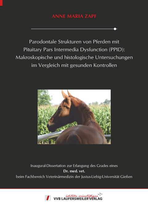
Parodontale Strukturen von Pferden mit Pituitary Pars Intermedia Dysfunction (PPID):
Makroskopische und histologische Untersuchungen im Vergleich mit gesunden Kontrollen
Seiten
2024
VVB Laufersweiler Verlag
9783835971707 (ISBN)
VVB Laufersweiler Verlag
9783835971707 (ISBN)
Pituitary Pars Intermedia Dysfunction (PPID) ist die häufigste neurodegenerative endokrine Erkrankung bei Pferden, die älter als 15 Jahre sind. Regelmäßig treten bei älteren Pferden Zahnerkrankungen und insbesondere Parodontitiden auf. Obwohl PPID bei geriatrischen Pferden und Zahnerkrankungen in allen Altersgruppen gut beschrieben sind, wurden mögliche Zusammenhänge zwischen dieser endokrinen Erkrankung und pathologischen Veränderungen der Zahnstrukturen von Pferden noch nicht hergestellt. Bei PPID sind Querverbindungen zu Immunfehlregulationen, gestörter Kollagensynthese sowie verzögerten Heilungsprozessen nachgewiesen. Daher war es das Ziel der publizierten Studien zunächst makroskopische und dann histologische Veränderungen der Gingiva und subgingivaler parodontaler Strukturen im Zusammenhang mit PPID zu identifizieren. Im ersten Teil der Studie wurden 14 Pferde mit klinischen Anzeichen von PPID und Adenom in der Pars intermedia der Hypophyse sowie 13 Kontrollpferde, die weder klinische Anzeichen noch PPID-assoziierte histologische Veränderungen in der Hypophyse zeigten, eingeschlossen. PPID-betroffene Pferde (26,9 ± 0,73 Jahre) waren signifikant älter als die Kontrollen (20,0 ± 1,24 Jahre; p = 0,0006). Erstmals wurden makroskopische Veränderungen der Gingiva bei Pferden mit PPID im Vergleich zu Kontrollen beschrieben. Das signifikant häufigere Auftreten von Veränderungen der Gingivastruktur mit einem voluminösen, unregelmäßigen Erscheinungsbild (p < 0,0001), ein unregelmäßiger Gingivaverlauf (p = 0,04) und vermehrt nachgewiesene vertiefte gingivale Sulci (p = 0,004) deuten auf eine Prädisposition von PPID-betroffenen Pferden für Parodontitis hin. Endokrinologische Pathomechanismen, die die geschwächte gingivale Gewebestruktur bei PPID erklären können, müssen künftig allerdings noch aufgezeigt werden.
Bei der zweiten Publikation handelt es sich um eine morphometrisch-deskriptive Fall-Kontroll-Studie. Es wurden insgesamt 145 Zahnlokalisationen von 10 PPID-betroffenen Pferden (27,3 ± 2,06 Jahre) mit 147 Zahnlokalisationen von 10 Kontrollpferden (21,4 ± 4,12 Jahre; p < 0,001) verglichen. Histologische Parameter waren Leukozyteninfiltration, Verhornung der Zahnfleischepithelien (orales Gingivaepithel, Sulkusepithel und Saumepithel), Blutgefäßversorgung des Parodontiums und Struktur des Zements. Die Verteilung und Lokalisation gingivaler Leukozyteninfiltrate bei PPID-betroffenen Pferden war häufiger multifokal bis konfluierend (p = 0,002) und reichte bis in tiefere Teile des Parodontiums, manchmal bis hinunter zum subgingivalen parodontalen Ligament (PDL). Ältere Tiere beider Gruppen zeigten eine höhere Prävalenz (PPID: OR 1,66; Kontrollen: OR 1,15) für eine hochgradige Leukozyteninfiltration im parodontalen Ligament. Das Alter war allerdings wichtiger als der PPID-Status für die stärkere Ansammlung und Anzahl der Leukozyteninfiltrate im PDL. Das Zahnzement an interdentalen Positionen zeigte viermal mehr Unregelmäßigkeiten bei PPID-betroffenen Pferden als bei Kontrollen, was für Futtereinspießungen, Diastematabildung und parodontale Erkrankungen prädisponiert. Es wurden zum ersten Mal an PPID erkrankte Pferde mit gesunden ≥ 15 Jahre alten Kontrollen in histologischen Parametern des equinen Parodontiums verglichen. Eine weitere Charakterisierung der Leukozyten wäre erforderlich, um ihre mögliche Rolle bei destruktiven parodontalen Prozessen bei PPID-betroffenen Pferden detailliert zu bewerten. Insgesamt lässt sich zusammenfassen, dass an PPID erkrankte Pferde im Vergleich zu gesunden Kontrollen sowohl makroskopisch als auch auf histologischer Ebene Veränderungen aufweisen, die für sich genommen prädisponierend für eine parodontale Erkrankung sein können. Insgesamt besitzt das Alter aber einen größeren Einfluss auf die Zähne und die parodontalen Strukturen des Pferdes als der PPID Status. Pituitary Pars Intermedia Dysfunction (PPID) is the most common neurodegenerative endocrine disease in horses older than 15 years. Dental diseases and especially periodontitis occur regularly in older horses. Although PPID in geriatric horses and dental disease in all age groups are well described, possible associations between this endocrine disorder and pathologic changes in equine dental structures have not yet been established. In the case of PPID, cross-connections to immune dysregulation, disturbed collagen synthesis and delayed healing processes have been proven. Therefore, the aim of the published studies was to first identify macroscopic and then histological changes in the gingiva and subgingival periodontal structures associated with PPID. In the first part of the study, 14 horses with clinical signs of PPID and adenoma in the pars intermedia of the pituitary gland and 13 control horses that showed neither clinical signs nor PPID-associated histological changes in the pituitary gland were included. PPID-affected horses (26.9 ± 0.73 years) were significantly older than controls (20.0 ± 1.24 years; p = 0.0006). For the first time, macroscopic changes in the gingiva of horses with PPID compared to controls were described. The significantly more frequent occurrence of changes in the gingival structure with a voluminous, irregular appearance (p < 0.0001), an irregular gingival course (p = 0.04) and increased evidence of deepened gingival sulci (p = 0.004) indicate a predisposition of PPID-affected horses for periodontitis. However, endocrinologically caused pathomechanisms that can explain the weakened gingival tissue structure in PPID still have to be identified in the future. The second publication is a morphometric-descriptive case-control study. A total of 145 tooth locations from 10 PPID-affected horses (27.3 ± 2.06 years) were compared with 147 tooth locations from 10 control horses (21.4 ± 4.12 years; p < 0.001). Histological parameters were leucocyte infiltration, keratinization of the gingival epithelium (oral gingival epithelium, sulcus epithelium and junctional epithelium), blood vessel supply to the periodontium and structure of the cementum. The distribution and localization of gingival leukocyte infiltrates in PPID-affected horses was more frequently multifocal to confluent (p = 0.002) and extended into deeper parts of the periodontium, sometimes down to the subgingival periodontal ligament (PDL). Older animals of both groups showed a higher prevalence (PPID: OR 1.66; controls: OR 1.15) of high-grade leukocyte infiltration in the periodontal ligament. However, age was more important than PPID status for the greater accumulation and number of leukocyte infiltrates in the PDL. The cementum at interdental positions showed four times more irregularities in PPID-affected horses than in controls, predisposing to food impaling, diastema formation and periodontal disease. For the first time horses suffering from PPID were compared with healthy old controls in histological parameters of the equine periodontium. Further characterization of leukocytes would be required to assess in detail their possible role in destructive periodontal processes in PPID-affected horses. Overall, it can be concluded that horses suffering from PPID show changes both macroscopically and histologically compared to healthy controls, which in themselves can be predisposing factor for periodontal disease. Overall, however, age has a greater influence than PPID status on the teeth and periodontal structures of the horse.
Bei der zweiten Publikation handelt es sich um eine morphometrisch-deskriptive Fall-Kontroll-Studie. Es wurden insgesamt 145 Zahnlokalisationen von 10 PPID-betroffenen Pferden (27,3 ± 2,06 Jahre) mit 147 Zahnlokalisationen von 10 Kontrollpferden (21,4 ± 4,12 Jahre; p < 0,001) verglichen. Histologische Parameter waren Leukozyteninfiltration, Verhornung der Zahnfleischepithelien (orales Gingivaepithel, Sulkusepithel und Saumepithel), Blutgefäßversorgung des Parodontiums und Struktur des Zements. Die Verteilung und Lokalisation gingivaler Leukozyteninfiltrate bei PPID-betroffenen Pferden war häufiger multifokal bis konfluierend (p = 0,002) und reichte bis in tiefere Teile des Parodontiums, manchmal bis hinunter zum subgingivalen parodontalen Ligament (PDL). Ältere Tiere beider Gruppen zeigten eine höhere Prävalenz (PPID: OR 1,66; Kontrollen: OR 1,15) für eine hochgradige Leukozyteninfiltration im parodontalen Ligament. Das Alter war allerdings wichtiger als der PPID-Status für die stärkere Ansammlung und Anzahl der Leukozyteninfiltrate im PDL. Das Zahnzement an interdentalen Positionen zeigte viermal mehr Unregelmäßigkeiten bei PPID-betroffenen Pferden als bei Kontrollen, was für Futtereinspießungen, Diastematabildung und parodontale Erkrankungen prädisponiert. Es wurden zum ersten Mal an PPID erkrankte Pferde mit gesunden ≥ 15 Jahre alten Kontrollen in histologischen Parametern des equinen Parodontiums verglichen. Eine weitere Charakterisierung der Leukozyten wäre erforderlich, um ihre mögliche Rolle bei destruktiven parodontalen Prozessen bei PPID-betroffenen Pferden detailliert zu bewerten. Insgesamt lässt sich zusammenfassen, dass an PPID erkrankte Pferde im Vergleich zu gesunden Kontrollen sowohl makroskopisch als auch auf histologischer Ebene Veränderungen aufweisen, die für sich genommen prädisponierend für eine parodontale Erkrankung sein können. Insgesamt besitzt das Alter aber einen größeren Einfluss auf die Zähne und die parodontalen Strukturen des Pferdes als der PPID Status. Pituitary Pars Intermedia Dysfunction (PPID) is the most common neurodegenerative endocrine disease in horses older than 15 years. Dental diseases and especially periodontitis occur regularly in older horses. Although PPID in geriatric horses and dental disease in all age groups are well described, possible associations between this endocrine disorder and pathologic changes in equine dental structures have not yet been established. In the case of PPID, cross-connections to immune dysregulation, disturbed collagen synthesis and delayed healing processes have been proven. Therefore, the aim of the published studies was to first identify macroscopic and then histological changes in the gingiva and subgingival periodontal structures associated with PPID. In the first part of the study, 14 horses with clinical signs of PPID and adenoma in the pars intermedia of the pituitary gland and 13 control horses that showed neither clinical signs nor PPID-associated histological changes in the pituitary gland were included. PPID-affected horses (26.9 ± 0.73 years) were significantly older than controls (20.0 ± 1.24 years; p = 0.0006). For the first time, macroscopic changes in the gingiva of horses with PPID compared to controls were described. The significantly more frequent occurrence of changes in the gingival structure with a voluminous, irregular appearance (p < 0.0001), an irregular gingival course (p = 0.04) and increased evidence of deepened gingival sulci (p = 0.004) indicate a predisposition of PPID-affected horses for periodontitis. However, endocrinologically caused pathomechanisms that can explain the weakened gingival tissue structure in PPID still have to be identified in the future. The second publication is a morphometric-descriptive case-control study. A total of 145 tooth locations from 10 PPID-affected horses (27.3 ± 2.06 years) were compared with 147 tooth locations from 10 control horses (21.4 ± 4.12 years; p < 0.001). Histological parameters were leucocyte infiltration, keratinization of the gingival epithelium (oral gingival epithelium, sulcus epithelium and junctional epithelium), blood vessel supply to the periodontium and structure of the cementum. The distribution and localization of gingival leukocyte infiltrates in PPID-affected horses was more frequently multifocal to confluent (p = 0.002) and extended into deeper parts of the periodontium, sometimes down to the subgingival periodontal ligament (PDL). Older animals of both groups showed a higher prevalence (PPID: OR 1.66; controls: OR 1.15) of high-grade leukocyte infiltration in the periodontal ligament. However, age was more important than PPID status for the greater accumulation and number of leukocyte infiltrates in the PDL. The cementum at interdental positions showed four times more irregularities in PPID-affected horses than in controls, predisposing to food impaling, diastema formation and periodontal disease. For the first time horses suffering from PPID were compared with healthy old controls in histological parameters of the equine periodontium. Further characterization of leukocytes would be required to assess in detail their possible role in destructive periodontal processes in PPID-affected horses. Overall, it can be concluded that horses suffering from PPID show changes both macroscopically and histologically compared to healthy controls, which in themselves can be predisposing factor for periodontal disease. Overall, however, age has a greater influence than PPID status on the teeth and periodontal structures of the horse.
| Erscheinungsdatum | 20.03.2024 |
|---|---|
| Reihe/Serie | Edition Scientifique |
| Verlagsort | Gießen |
| Sprache | deutsch |
| Maße | 148 x 210 mm |
| Gewicht | 200 g |
| Themenwelt | Veterinärmedizin ► Allgemein |
| Schlagworte | Cushing-Syndrom • Endokrinopathie beim Pferd • Equines • metabolische Erkrankung • Neurologie • Pferd |
| ISBN-13 | 9783835971707 / 9783835971707 |
| Zustand | Neuware |
| Informationen gemäß Produktsicherheitsverordnung (GPSR) | |
| Haben Sie eine Frage zum Produkt? |
Mehr entdecken
aus dem Bereich
aus dem Bereich
Buch | Softcover (2025)
Mensch & Buch (Verlag)
CHF 69,85


