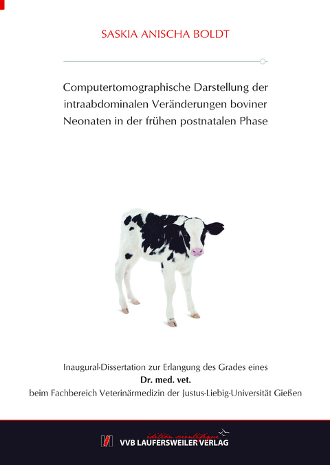
Computertomographische Darstellung der intraabdominalen Veränderungen boviner Neonaten in der frühen postnatalen Phase
Seiten
2023
VVB Laufersweiler Verlag
978-3-8359-7139-4 (ISBN)
VVB Laufersweiler Verlag
978-3-8359-7139-4 (ISBN)
- Keine Verlagsinformationen verfügbar
- Artikel merken
Ziel dieser Arbeit war die physiologisch-topographischen Verhältnisse der abdominalen Organe des neugeborenen Kalbes darzustellen. Im Mittelpunkt dieser Untersuchung standen die Magenabteilungen, der Darm, die Leber und Nieren.
Zu diesem Zweck wurden von 15 klinisch gesunden Kälbern der Rasse Holstein Friesian computertomographische Scans durchgeführt. Diese Aufnahmen fanden am 1., 7., 14. und 21. Lebenstag statt. Am ersten Lebenstag der Kälber entstanden drei Aufnahmen in einem Abstand von sechs Stunden. Die erste Untersuchung der Kälber fand direkt nach der Geburt statt.
Folgende relevante Ergebnisse wurden erzielt:
-Bezogen auf das Gesamtmagenvolumen besitzt das Retikulorumen einen durchschnittlichen Anteil von 50,1 %, der Labmagen nimmt ca. 42 % des Volumens direkt nach der Geburt ein.
-Zwölf Stunden p. n. und nach der Aufnahme von Kolostrum steigt das Volumen des Labmagens auf 1,6 l und überragt das Retikulorumen um mehr als das doppelte. Somit ändern sich auch die prozentualen Volumenanteile. Pansen und Retikulum beanspruchen nur noch 28,2 % und der Labmagen 69 % des Gesamtmagenvolumens.
-Das Volumenmaximum erreicht der Labmagen eine Woche p. n. und sinkt im weiteren Verlauf durch den Beginn der Rohfaserfütterung. Im gleichen Zuge steigt das Volumen des Retikulorumens stetig an und nimmt prozentual am Gesamtmagenvolumen zu.
-Eine deutliche Pansenschichtung konnte bei 20 % der Kälber eine Woche p. n. und nach 21 Tagen bei allen Kälbern nachgewiesen werden. Die Untersuchungen am ersten Lebenstag des Kalbes zeigen eine Zweischichtung des Pansens aus luftähnlichem und homogenem Inhalt. Der homogene ventrale Inhalt setzt sich nach der Kolostrumverabreichung vor allem aus diesem und dem Kaseinkuchen zusammen.
-Es konnte eine fortschreitende Parenchymverdichtung der Leber in den ersten drei Lebenswochen festgestellt werden. Bei den Untersuchungen am Geburtstag wiesen die Kälber zentral durchschnittliche Werte von 51,54 bis 55,97 HU und peripher von 56,98 bis 59,85. In den folgenden drei Wochen stieg die mittlere Dichte sowohl peripher als auch zentral auf Werte von 61,26 HU (zentral) und 67,29 HU (peripher) an.
-Es zeigt sich eine Volumenveränderung der Leber am ersten Lebenstag. Das hepatische Gewebevolumen nahm bei allen Kälbern in den ersten Lebensstunden signifikant ab. Eine Stunde p. n. ergaben die Messungen ein durchschnittliches Volumen von 1003,4 cm³, welches sich zur zweiten Untersuchung auf 892,3 cm³ und zur dritten auf 887,1 cm³ verringerte.
-Die Nieren zeigten über den Untersuchungsverlauf keine signifikanten topographischen Lageveränderungen. Bereits zur ersten Untersuchung befindet sich die linke Niere deutlich kaudal der rechten. Die linke Niere befand sich in den ersten drei Lebenswochen des Kalbes links der Medianen und lag der Bauchwand dorsolateral an oder wurde vom Pansen in Richtung der Medianen gedrängt.
-Der Hilus der linken Niere zeigt im kaudalen Bereich nach ventral, im kranialen Bereich zur Medianen. Diese 90° Rotation lässt sich bereits eine Stunde post natum darstellen.
-Die kraniokaudale Länge der Nieren verändert sich in den ersten drei Lebenswochen nicht signifikant, dafür nimmt das Volumen innerhalb der ersten Lebenswoche um 31 % zu und erreicht am 21. Lebenstag eine Zunahme im Vergleich zur initialen Situation um 50%.
Die vorliegende Studie zeigt erstmals die abdominalen topographischen Verhältnisse beim neugeborenen Kalb mittels Computertomographie in einer hohen Untersuchungsfrequenz am ersten Lebenstag. Sie stellt damit die Grundlage für weitere Untersuchungen, z. B. im Zusammenhang mit der Fragestellung des Drenchens, dar. The aim of this study was to describe the relationships of the abdominal organs of the newborn calf regarding its physiology and topography. The focus of this investigation was on the stomach departments, the intestine, the liver and kidneys. For this purpose, computerized tomographic scans were performed on 15 healthy Holstein Friesian calves. Imaging was conducted on the 1st, 7th, 14th and 21st day of life. On the first day of life, an examination of the calves was performed right after birth and three shots were taken every six hours.
In summary following relevant results were obtained:
-Based on the total stomach volume, the reticulum volume has an average share of 50.1 %, the abomasum occupies about 42 % of the volume immediately after birth.
-12 hours post natum and after ingestion of colostrum, the volume of the abomasum increases to 1.6 l and exceeds the reticulum volume by more than twice. It can be stated that total volumes of abomasum and reticulum change drastically after the first ingestion of milk. Rumen and reticulum require only 28.2 % and the abomasum 69 % of the total stomach volume.
-The abomasal volume reaches its maximum one-week post natum and decreases with the beginning of first fibre intake. At the same time, the volume of the reticulum increases steadily in its total volume and percentage among the total stomach volume.
-Distinct rumen stratification was observed in 20 % of calves one-week post natum, after 21 days this could be detected in all calves. The investigations of one-day-old calves show a two-layered rumen of air-like and homogeneous contents. The homogeneous ventral content is composed after first milk administration, resulting in a mixture of colostrum and incurred casein cake.
-Concerning the liver, a steady parenchymal compression in the first three weeks of life could be observed. In the scans from the first day the calves central average values of 51.54 to 55.97 HU and peripherally 56.98 to 59.85 HU were measured. In the following three weeks, the mean density increased both peripherally and centrally to levels of 61.26 HU and 67.29 HU.
-Moreover, a change in total liver volume on the first day of life could be quantified. The hepatic tissue volume decreased significantly in all calves in the first few hours of life. One-hour post natum the measurements indicated an average volume of 1003.4 cm³, which decreased to 892.3 cm³ for the second study and to 887.1 cm³ for the second.
-The kidneys showed no significant topographical changes in the course of the study. Already for the first examination, the left kidney is indistinctively located caudal to the right. The left kidney was located to the left of the median in the first three weeks of life of the calf and dorsolateral to the abdominal wall or pushed by the rumen towards the median.
-The hilum of the left kidney points ventrally in the caudal region and the cranial region medially. This 90° rotation due to the strain of the rumen can already be observed one-hour post natum.
-The craniocaudal length of the kidneys does not change significantly in the first three weeks of life. Nevertheless, the volume increases within the first week of life by 31 %. It reaches on the 21st day of life a increase compared to the initial situation by 50 %.
The present study shows the abdominal topographic conditions in neonatal calves using computed tomography at a high examination frequency for the first time. It thus provides the basis for further investigations, e.g. in connection with the question of drenching.
| Erscheinungsdatum | 11.08.2023 |
|---|---|
| Reihe/Serie | Edition Scientifique |
| Verlagsort | Gießen |
| Sprache | deutsch |
| Maße | 148 x 215 mm |
| Gewicht | 330 g |
| Themenwelt | Veterinärmedizin ► Allgemein |
| Veterinärmedizin ► Großtier | |
| Schlagworte | CT • Frischgeborene Tiere • Tiergeburten |
| ISBN-10 | 3-8359-7139-5 / 3835971395 |
| ISBN-13 | 978-3-8359-7139-4 / 9783835971394 |
| Zustand | Neuware |
| Informationen gemäß Produktsicherheitsverordnung (GPSR) | |
| Haben Sie eine Frage zum Produkt? |

