Der Einfluss von HIF 1α auf die Immunantwort des Wirts bei Chlamydien-Infektionen des weiblichen Genitaltrakts der Maus
Seiten
2023
VVB Laufersweiler Verlag
978-3-8359-7099-1 (ISBN)
VVB Laufersweiler Verlag
978-3-8359-7099-1 (ISBN)
- Keine Verlagsinformationen verfügbar
- Artikel merken
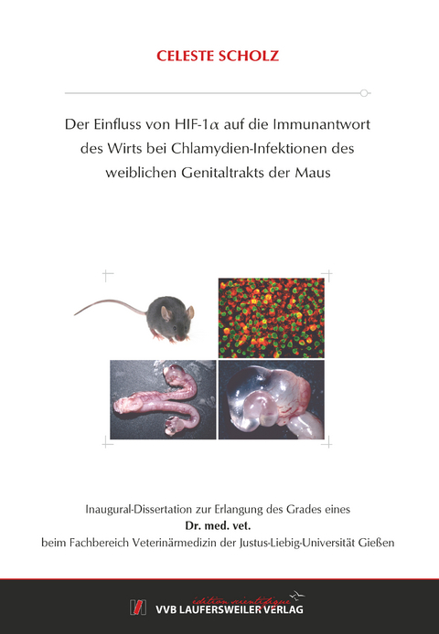
Chlamydia trachomatis (C. trachomatis) ist ein obligat intrazelluläres Bakterium, das die häufigste sexuell übertragene bakterielle Infektion des Menschen weltweit verursacht. Wenn solche Infektionen unentdeckt bleiben, können sie bei Frauen schwere genitale Erkrankungen auslösen, die im schlimmsten Fall zu Unfruchtbarkeit führen. Aus ethischen Gründen sind Studien an Menschen nur beschränkt möglich, weshalb Mausmodelle zur Untersuchung von Chlamydien-Infektionen verwendet werden können. C. trachomatis und C. muridarum sind sich genetisch sehr ähnlich, weisen ca. 90 % Orthologie des Gengehalts auf und gehören der selben Gattung an. Da humane C. trachomatis- und murine C. muridarum-Infektionen klinisch ähnlich verlaufen, eignet sich das Progesteron-abhängige C. muridarum-Mausmodell als Modellsystem für humane C. trachomatis-Infektionen. Bei Mäusen wie bei Menschen steigen die Chlamydien nach vaginaler Infektion über Zervix, Uterus und Tuben bis zu den Ovarien auf. Dort verursachen sie beim Menschen Verklebungen an den Tuben und bei Mäusen Hydrosalpingen, die aus serösen Einschlüssen bestehen. Beide Pathologievarianten lösen bei den jeweiligen Individuen im schlimmsten Fall eine Infertilität aus. Die Umgebungsbedingungen sind maßgeblich für die Entstehung und Progression von Entzündungen, weshalb das hypoxische Niveau des Genitaltrakts bei Chlamydien-Infektionen eine wichtige Rolle spielt. Hypoxische Reaktionen in Gewebe und Zellen werden unter anderem durch Transkriptionsfaktoren wie den Hypoxie-Induzierbaren-Faktor- (HIF-) 1α vermittelt, der nach Aktivierung über metabolische Veränderungen und zelluläre Anpassungen in die Immunantwort des Wirts eingreift. Es wurde ein etabliertes, Progesteron-abhängiges C57BL/6-Mausmodell durch ein HIF 1α-Knockout-Mausmodell erweitert. Die Deletion von HIF 1α bezog sich auf myeloide Zellen und die Mäuse besaßen einen C57BL/6 Hintergrund. Mittels dieses Mausmodells wurde der Einfluss von HIF 1α auf die vaginale C. muridarum-Besiedlung, die Entstehung von Ovarialpathologien und die Immunantwort auf vaginale C. muridarum-Infektionen untersucht.
Nach vaginaler C. muridarum-Infektion war zu Beginn der akuten Infektion bei den HIF 1α / -Mäusen eine geringere vaginale Chlamydien-Last im Vergleich zu den Wildtyp- (WT)-Tieren sichtbar. Dies wurde mittels Wiederanzuchtassays der vaginal abgestrichenen infektiösen C. muridarum-Einheiten ermittelt. Durchflusszytometrisch wurde die Menge der lokalen Immunzellen im Uterus der Mauslinien ermittelt. Dabei konnten Unterschiede zwischen den Genotypen ohne statistische Signifikanz in der Menge der Makrophagen, Granulozyten, CD4+- und CD8+-T-Zellen gezeigt werden. In Form von makroskopisch und mikroskopisch sichtbaren Hydrosalpingen beschleunigte das Fehlen von HIF 1α die Entwicklung von Pathologien an Tag 21 p. i. signifikant bei den HIF 1α-/--Mäusen im Vergleich zu den WT-Kontrollen. An Tag 43 p. i. nivellierte sich dieser Unterschied zwischen den Genotypen. Nach Messung der C. muridarum-spezifischen Serum-IgG-Titer mittels ELISA war ein signifikanter Unterschied zwischen den Mauslinien an Tag 21 und 43 p. i. sichtbar, der in einer Verringerung der IgG-Level bei den HIF 1α / -Mäusen im Vergleich zu den WT-Tieren bestand. Die Zusammenhänge zwischen vaginaler Chlamydien-Besiedlung, den zellulären Veränderungen des lokalen Immunsystems, der Entwicklung von Ovarialpathologien und der systemischen C. muridarum-spezifischen IgG-Antikörperbildung mit HIF 1α werden in der vorliegenden Arbeit diskutiert und interpretiert.
Zusammenfassend hat HIF 1α einen signifikanten Einfluss auf die Entwicklung von Ovarialpathologien nach Chlamydien-Infektionen und auf die Entstehung von C. muridarum-spezifischem IgG im Blutserum. Dies ist möglicherweise auf eine Kompensation einer eingeschränkten Funktionalität von HIF 1α defizienten Immunzellen durch eine Erhöhung der Menge von Makrophagen und Granulozyten im Uterus zurückzuführen. Im Weiteren könnte diese Zellerhöhung zu einer vermehrten Gewebszell-Proliferation geführt haben, die signifikant steigernd auf die Entwicklung von Ovarialpathologien bei den HIF 1α / -Mäusen wirkte. Die signifikante Verringerung der C. muridarum-spezifischen IgG-Antwort im Vergleich zu den WT-Kontrolltieren kam womöglich dosisabhängig zustande, da durch das Fehlen von HIF 1α geringere Chlamydien-Infektionen bei den Knockout-Tieren vorherrschten und sie daher möglicherweise geringere IgG-Antworten ausbildeten als die WT-Kontrolltiere. Klinisch ist vor allem interessant, dass die Stärke der Chlamydien-Infektion und die Verweildauer der Chlamydien im Genitaltrakt der Mäuse einen Einfluss auf die Ausbildung von Pathologien besitzen könnten. Womöglich könnte die Pathologieentwicklung durch die Ausprägung der innaten und adaptiven Immunantworten der Mäuse bedingt gewesen sein. Chlamydia trachomatis (C. trachomatis) is an obligate intracellular bacterium that causes the most common sexually transmitted bacterial infection in humans worldwide. If undetected, such infections can cause severe genital diseases in women, which in the worst cases can lead to infertility. For ethical reasons, there are limitations in human studies, which is why mouse models can be used to study chlamydial infections. C. trachomatis and C. muridarum are genetically very similar, show about 90 % orthology of gene contents and belong to the same genus. Since human C. trachomatis and murine C. muridarum infections are clinically comparable and can lead to infertility in the end, a progesterone dependent C. muridarum mouse model is a suitable and well-established model system for human C. trachomatis infections. In mice as in humans, chlamydiae ascend after vaginal infection via the cervix, uterus and fallopian tubes to the ovaries. They can cause adhesions in human fallopian tubes and hydrosalpinges, which consist of serous inclusions, in mice. At worst, both pathology variants can cause infertility in the respective individuals. Considering that the environmental conditions are decisive for the development and progression of inflammations, the hypoxic environment present in the genital tract plays an important role in genital chlamydial infections. Hypoxic reactions in tissue and cells are mediated by transcription factors such as the Hypoxia Inducible Factor (HIF) 1α. After activation, HIF 1α interferes with the host's immune response via metabolic changes and cellular adaptations. In the present work, an established progesterone dependent C57BL/6 mouse model was extended by a HIF 1α knockout mouse model. The deletion of HIF 1α was related to myeloid cells and mice had a C57BL/6 background. Using this mouse model, the impact of HIF 1α on the vaginal chlamydial load, on the pathology formation at the ovaries and on the immune response of vaginal C. muridarum infections was investigated.
After vaginal C. muridarum infection, a lower vaginal chlamydial load was detectable in the HIF 1α / mice compared to the wildtype (WT) animals at the beginning of the acute infection. This was determined using recovery assays of vaginally swabbed infective progeny of C. muridarum. The amount of local immune cells in the uterus of the mouse lines was determined by flow cytometry. Differences between the genotypes without statistical significance in the number of macrophages, granulocytes, CD4+- and CD8+-T cells could be shown. The absence of HIF 1α significantly accelerated the formation of macroscopically and microscopically visible hydrosalpinges at day 21 p. i. in the HIF 1α / mice compared to the WT controls. On day 43 p. i. this difference between the genotypes leveled out. After evaluating the C. muridarum specific Serum IgG levels in the blood serum of mice via ELISA, a significant difference between the mouse lines on day 21 and 43 p. i. was visible. It consisted in a reduction of the IgG levels in the HIF 1α / mice compared to the WT animals. The correlations between vaginal chlamydial colonization, the cellular changes of the local immune system, the development of ovarian pathologies and the systemic IgG antibody formation with HIF 1α are discussed and interpreted in the present work.
Concluding, HIF 1α has a significant impact on the development of ovarian pathologies after chlamydial infections and on the formation of C. muridarum specific IgG in the blood serum. This could be due to a compensation of a reduced efficiency of HIF 1α deficient immune cells by an increase in the number of macrophages and granulocytes in the uterus. Furthermore, this cell increase could have led to increased tissue cell proliferation, which had a significantly increasing effect on the development of ovarian pathologies in the HIF 1α / mice. The significant reduction in the C. muridarum specific IgG response compared to the WT control animals was probably dose dependent, since the lack of HIF 1α resulted in lower chlamydial colonization in the knockout animals. Therefore, these mice developed lower IgG responses than the WT control animals. From a clinical point of view, it is of particular interest that the severity of the chlamydia infection and the length of time the chlamydia remain in the genital tract of the mice could have an impact on the development of pathologies. The development of the pathology could possibly have been caused by the expression of the innate and adaptive immune responses of the mice.
| Erscheinungsdatum | 16.03.2023 |
|---|---|
| Reihe/Serie | Edition Scientifique |
| Verlagsort | Gießen |
| Sprache | deutsch |
| Maße | 148 x 210 mm |
| Gewicht | 180 g |
| Themenwelt | Veterinärmedizin ► Allgemein |
| Veterinärmedizin ► Klinische Fächer ► Parasitologie | |
| Veterinärmedizin ► Klinische Fächer ► Pharmakologie / Toxikologie | |
| Schlagworte | Bakterium • Chlamydien • Erreger • HIF 1a • Immunantwort |
| ISBN-10 | 3-8359-7099-2 / 3835970992 |
| ISBN-13 | 978-3-8359-7099-1 / 9783835970991 |
| Zustand | Neuware |
| Informationen gemäß Produktsicherheitsverordnung (GPSR) | |
| Haben Sie eine Frage zum Produkt? |
Mehr entdecken
aus dem Bereich
aus dem Bereich
Buch | Spiralbindung (2023)
Schlütersche (Verlag)
CHF 249,95


