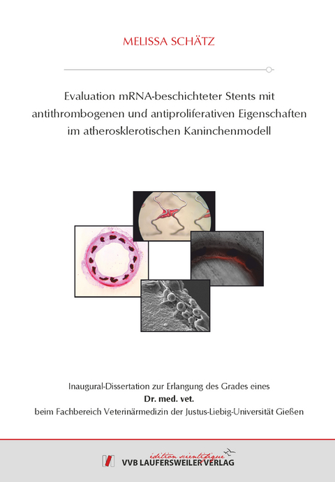
Evaluation mRNA-beschichteter Stents mit antithrombogenen und antiproliferativen Eigenschaften im atherosklerotischen Kaninchenmodell
Seiten
2021
VVB Laufersweiler Verlag
978-3-8359-6972-8 (ISBN)
VVB Laufersweiler Verlag
978-3-8359-6972-8 (ISBN)
- Keine Verlagsinformationen verfügbar
- Artikel merken
Aufgrund der hohen Prävalenz und der häufigen Todesfolge sind kardiovaskuläre Erkrankungen beim Menschen weltweit von großer Bedeutung. Dabei stellt die perkutane transluminale Koronarangioplastie, bei welcher Stents als Gefäßstützen in die geschädigten Gefäße implantiert werden, eine wichtige Behandlungsmöglichkeit dar. Trotz zahlreicher Fortschritte in Stentdesign und Wirkstoffbeschichtung kommt es immer wieder zu schweren thrombotischen Komplikationen und zum erneuten Verschluss des betroffenen Gefäßes.
Einen vielversprechenden Ansatz zur Reduktion der genannten Komplikationen bietet das Protein CD39, welches durch die Hydrolyse von ADP und ATP sowohl die Aktivierung der Thrombozyten als auch die Proliferation glatter Muskelzellen inhibiert. Durch die Beschichtung von Stents mit in vitro generierter CD39 mRNA soll eine lokale Protein-Überexpression am betroffenen Gefäßabschnitt induziert werden, um die positiven Effekte des Proteins zu nutzen. In der vorliegenden Arbeit wurde deshalb sowohl die Transfektionseffizienz sowie die Funktionalität des translatierten CD39 Proteins als auch die Hämokompatibilität der mit CD39 mRNA beschichteten Stents in vitro untersucht und anschließend die antithrombogene und antiproliferative Eigenschaft im atherosklerotischen In-vivo-Kaninchenmodell evaluiert.
Zunächst wurden HEK-Zellen mit unterschiedlichen Varianten der CD39 mRNA transfiziert und nach einem Zeitraum von 24, 48 und 72 Stunden die Expression des Proteins und die Aktivierung der Thrombozyten mittels Durchflusszytometrie analysiert. Mit Hilfe des Malachite Green Phosphate Assay Kits wurde zudem die Funktion des Proteins durch die Messung des freien Phosphats überprüft. Es zeigte sich, dass verschiedene Varianten der synthetisch hergestellten CD39 mRNA teilweise deutliche Unterschiede in der Expression und der Funktionsfähigkeit des Proteins aufweisen. Die stärkste Expression und Funktionalität wurde dabei beim Wildtyp und den Varianten optimiert und 49 nachgewiesen.
Darüber hinaus konnte durch die Inkubation unterschiedlich beschichteter Stents (CD39 mRNA, BMS, Sirolimus) mit humanem Blut in einem Agitations-Modell die Hämokompatibilität der CD39 mRNA-Beschichtung gezeigt werden. Dabei wiesen die mit CD39 mRNA beschichteten Stents eine den BMS und Sirolimus-Stents vergleichbare, geringgradige thromboadhäsive Wirkung auf.
Für die In-vivo-Evaluation wurden die CD39 mRNA beschichteten Stents sowie verschiedene Kontrollgruppen in die Carotiden von New-Zealand-White Kaninchen implantiert. Nach 3, 14 und 30 Tagen wurden die Kaninchen euthanasiert und die Blutgefäße mit den enthaltenen Stents entnommen. Dabei konnte die Expression des Proteins CD39 drei Tage nach der Implantation erfolgreich nachgewiesen werden. Nach 14 bzw. 30 Tagen wurde ausschließlich bei einem Präparat eine verstärkte Expression festgestellt. Die Freisetzung der mRNA sowie die anschließende Expression des Proteins in vivo ist also grundsätzlich auch nach einem längeren Zeitraum möglich, jedoch war dieses Ergebnis in der vorliegenden Arbeit nicht reproduzierbar. Darüber hinaus zeigte sich in allen Gruppen, dass die Stärke der Neointimabildung und damit der Grad der Stenose sowohl innerhalb eines einzelnen Präparates als auch innerhalb einer Gruppe stark variierte. Es konnte kein signifikanter Unterschied zwischen der Stenoserate der einzelnen Gruppen festgestellt werden. Ein gewisser antithrombotischer Effekt ist dagegen erkennbar, denn im Gegensatz zu den übrigen Gruppen konnte bei einer Gruppe mit CD39 mRNA beschichteten Stents (CD39 Coating 2) keine Thrombenbildung nachgewiesen werden.
Aufgrund der geringen Probenzahl und der starken Varianz innerhalb der meisten Gruppen ist eine fundierte Aussage nicht möglich. Für statistisch signifikante Werte wird eine höhere Probenzahl benötigt, weshalb weiterführende Untersuchungen zur CD39 mRNA- Beschichtung der Stents erforderlich sind. Dabei sollte insbesondere die Freisetzungskinetik der Matrix sowie die Konzentration der mRNA überprüft und gegebenenfalls angepasst werden, um die Freisetzung der mRNA über einen längeren Zeitraum sicherzustellen. Darüber hinaus erscheint die Analyse einer Kombination von CD39 mRNA mit weiteren Substanzen vielversprechend, um die thromboadhäsive Wirkung der Beschichtung weiter zu senken und den antiproliferativen Effekt der mRNA zu unterstützen. Zusätzlich sollte für weitere Untersuchungen eine weniger invasive, alternative Operationsmethode in Betracht gezogen werden, um die lokale Manipulation am Ort der Implantation zu verringern und damit die Vergleichbarkeit der Ergebnisse zu verbessern.
Zusammenfassend gab es Hinweise auf die Expression der CD39 mRNA im betroffenen Gefäßabschnitt. Es zeigte sich, dass die CD39 mRNA-Beschichtung insgesamt mit den bereits etablierten BMS und Sirolimus-Stents vergleichbar ist. Dabei besteht durchaus ein gewisses Potenzial, die Thrombenbildung innerhalb eines Stents zu verringern und darüber hinaus die Neointimabildung am betroffenen Gefäßabschnitt, und damit die Gefahr der Restenose, zu reduzieren. Um diese Tendenz weiter zu untermauern und die Effekte zu validieren, sind weitere Untersuchungen notwendig. Insbesondere sollte dabei ein verbesserter Tierversuchsaufbau mit höheren Fallzahlen verwendet werden, um die therapeutischen Effekte besser herausarbeiten zu können. Möglicherweise kann z.B. durch die Kombination von CD39 mRNA mit synergistischen Substanzen das Risiko der Restenose und Thrombose nach einer Stentimplantation reduziert werden, um Patienten längerfristig vor schwerwiegenden Komplikationen zu schützen. Cardiovascular diseases are of global significance due to their high prevalence and are a worldwide leading cause of death. One of the most important methods of treatment is the percutaneous coronary intervention in which stents are used to stabilize the affected vessel section. Despite several improvements in stent design and stent coatings there are often still serious thrombotic complications and restenosis.
The protein CD39 shows promise to reduce these complications: By hydrolyzing ADP and ATP, the activation of platelets and the proliferation of the smooth muscle cells could be inhibited. Using in vitro generated CD39 mRNA as a new stent coating should induce a local overexpression of this protein at the affected vessel section thereby displaying the above mentioned positive effects of CD39. In the current study we examined the transfection efficiency and function of CD39 mRNA as well as the haemocompatibility of CD39 mRNA coated stents followed by the evaluation of the antithrombotic and antiproliferative effects in an atherosclerotic in vivo rabbit model.
First, HEK cells were transfected with five different types of CD39 mRNA. After 24, 48 and 72 hours the expression of the protein and the activation of human platelets were analyzed with flow cytometry. Additionally, the function of the protein was tested by measuring the free phosphate using the Malachite Green Phosphate Assay. It reveals that different variations of the synthetically produced CD39 mRNA showed differences in the expression and the functionality of the given protein. The three types “Wildtyp”, “optimiert” und “49” showed the strongest expression and function.
Furthermore, it was possible to demonstrate the haemocompatibility of the CD39 mRNA coating by incubating different coated stents (CD39 mRNA, BMS, Sirolimus) with human blood using an agitation model. Overall, the CD39 mRNA coated stents possessed a comparable, marginal thromboadhesive effect in comparison to BMS or sirolimus coated stents.
For the in vivo evaluation, the CD39 mRNA coated stents and different control groups were implanted in the carotids from New-Zealand-White rabbits. After 3, 14 and 30 days the rabbits were euthanized and the vessels containing the stents were explanted. The expression of the protein CD39 could be successfully demonstrated three days after implantation. After 14 and 30 days the expression could only be detected in one sample. Generally, the release of the mRNA and the following expression of the protein in vivo are possible, but this result could not be reproduced in the presented study. Moreover, another result of this examination was that the amount of neointima formation varied strongly within one preparation and among one group. There was also no significant difference in the degree of stenosis of the individual groups. However, a certain antithrombotic effect is visible, because in contrast to the other groups there was one CD39 mRNA coated group (CD39 Coating 2) without any thrombus formation.
Because of the small number of samples and the strong variance within most of the given groups it is impossible to make a funded statement. To gain statistically significant statements, higher numbers of samples should be used. Further studies of the introduced stent coating are necessary. Thereby the release kinetics of the matrix and the concentration of the mRNA should be proven and if applicable adapted to guarantee the release of the mRNA over an extended period. Moreover, it seems promising to analyze the combination of CD39 mRNA with other substances to reduce the thromboadhesive effect of the coating even further and to support the antiproliferative effect of the mRNA. In addition, consideration should be given to an alternative operative procedure to decrease local manipulation at the implantation site to improve the comparability between the results.
In summary there are indications for the expression of CD39 mRNA in the affected tissue section. It was shown that the new, biodegradable CD39 mRNA coating is comparable with the already established BMS and sirolimus coated stents. It offers the potential to reduce thrombus formation within a stent and furthermore may decrease the risk of restenosis. To confirm this tendency and to validate these effects further research is necessary. Particularly the structure of the animal studies should be improved and the number of samples should increase to show therapeutic effects. For example it might be possible to reduce the risk of restenosis and thrombosis after stent implantation with a combination of CD39 mRNA and synergistic substances to protect patients from serious long-term complications.
| Erscheinungsdatum | 31.01.2022 |
|---|---|
| Reihe/Serie | Edition Scientifique |
| Verlagsort | Gießen |
| Sprache | deutsch |
| Maße | 148 x 210 mm |
| Gewicht | 180 g |
| Themenwelt | Veterinärmedizin ► Allgemein |
| Schlagworte | Herz • mRNA • Verkalkung |
| ISBN-10 | 3-8359-6972-2 / 3835969722 |
| ISBN-13 | 978-3-8359-6972-8 / 9783835969728 |
| Zustand | Neuware |
| Haben Sie eine Frage zum Produkt? |

