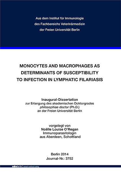
Monocytes and macrophages as determinants of susceptibility to infection in lymphatic filariasis
Seiten
2015
|
1. Aufl.
Mensch & Buch (Verlag)
978-3-86387-584-8 (ISBN)
Mensch & Buch (Verlag)
978-3-86387-584-8 (ISBN)
- Keine Verlagsinformationen verfügbar
- Artikel merken
BACKGROUND & AIMS: Helminths induce strong regulatory and T helper 2-type responses by targeting host cells. In lymphatic filariasis, the host response to this, together with a multitude of other factors, determines whether a person remains infection and disease free or develops a successful infection. Infection results in either asymptomatic infection, which benefits transmission of the parasite, or chronic pathology, which is responsible of the high levels of morbidity seen in patients. Monocytes and macrophages contribute to helminthinduced dysfunction of the immune response through modulation by microfilariae in the blood and tissues. During patent infection monocytes encounter microfilariae in the blood, an event that occurs in asymptomatically infected patients who are immunologically hyporeactive. Furthermore helminths induce regulatory antibody responses that may impact on disease outcome. Other disease models have shown that altered glycosylation of the IgG Fc region correlates with pathology, whereby decreased galactosylation is associated with inflammation and increased sialylation is associated with anti-inflammatory responses. The aim of this project was to determine whether microfilariae act on blood monocytes and macrophages to induce a regulatory phenotype that interferes with innate and adaptive responses. Furthermore the IgG glycosylation profile of the different disease outcomes was compared with determine a role for glycosylation in lymphatic filariasis.
PRINCIPAL FINDINGS: Monocytes and in vitro generated macrophages from filaria nonendemic normal donors stimulated with Brugia malayi microfilarial (Mf) lysate but not adult female lysate show a drastically altered phenotype. Monocytes stimulated with Mf lysate develop a defined regulatory phenotype, characterised by expression of IL-10 and PD-L1. Importantly, this regulatory phenotype was recapitulated in monocytes from Wuchereria bancrofti asymptomatically infected individuals but not patients with pathology or endemic normals. Monocytes from non-endemic donors stimulated with Mf lysate directly inhibited CD4+ T cell proliferation and cytokine production. CD4+ T cell IFN-γ responses were restored by neutralising IL-10 or PD-1. Furthermore, macrophages stimulated with Mf lysate expressed high levels of IL-10 and had suppressed phagocytic abilities. Finally Mf lysate applied during macrophage differentiation in vitro selectively interfered with macrophage abilities to respond to LPS stimulation. Additionally, Fc region N-linked glycans of total IgG from W. bancrofti-exposed donors were analysed. Using capillary electrophoresis it was found that there was no difference in galactosylation of total IgG between the different disease outcomes, however, asymptomatically infected patients had significantly lower levels of disialylated IgG compared with endemic normals and patients with pathology.
CONCLUSIONS & SIGNIFICANCE: Conclusively, this study demonstrates that Mf lysate stimulation of monocytes from healthy donors in vitro induces a regulatory phenotype, able to interfere with CD4+ T cell responses. This phenotype is directly reflected in monocytes from filarial patients with asymptomatic infection but not patients with pathology or endemic normals. The results suggest that suppression of T cell functions typically seen in lymphatic filariasis is caused by microfilaria-modulated monocytes in an IL-10- or PD-1-dependent manner. Together with suppression of macrophage innate responses, this may contribute to the overall down-regulation of immune responses observed in asymptomatically infected patients. Die Rolle von Monozyten und Makrophagen in der Ausprägung der Empfänglichkeit für Infektionen mit lymphatischer Filariose
HINTERGRUND UND ZIEL DER ARBEIT:
Helminthen induzieren starke regulatorische und Thelfer 2 Immunantworten, indem Wirtszellen gezielt moduliert werden. Die Immunantwort des Wirtes gegen Erreger der lymphatischen Filariose entscheidet darüber, ob ein Individuum resistent ist oder erfolgreich infiziert wird. Darüberhinaus führt die Infektion entweder zu einer klinisch asymptomatischen Manifestation, welche die Transmission des Parasiten begünstigt oder zu chronischer Pathologie, welche für die hohe Morbidität von lymphatischer Filariose-Patienten verantwortlich ist. Monozyten und Makrophagen tragen zur helminthen-induzierten Dysfunktionalität der Immunantwort bei, indem sie von Mikrofilarien in Blut und Gewebe moduliert werden. Während einer patenten Infektion treffen Monozyten im Blut auf zirkulierende Mikrofilarien ausschliesslich in asymptomatischen Patienten, welche sich durch eine Hyporeaktivität ihrer Immunantwort auszeichnen. Weiterhin induzieren Helminthen regulatorische Antikörperisotypen, welche einen Einfluss auf den Verlauf der Infektion haben. Aus anderen Krankheitsmodellen ist zudem bekannt, dass eine veränderte Glykosylierung der IgG Fc Region mit einer Pathologie einhergeht. Das Ziel dieser Arbeit war zu untersuchen, ob Mikrofilarien Monozyten aus dem Blut oder Makrophagen zu einem regulatorischen Phänotyp induzieren können, welche modulierte angeborene und adaptiven Immunantworten vermitteln. Weiterhin sollte das IgG Glykosylierungsprofil von Wuchereria bancrofti infizierten Individuen bestimmt werden, um einen eventuellen Einfluss der Glykosylierung auf das Krankheitsbild zu ermitteln.
ERGEBNISSE: Monozyten und in vitro generierte Makrophagen von non-endemischen Individuen zeigen einen drastisch veränderten Phänotyp nach Stimulation mit Mikrofilarien (Mf) Lysat; adultes Weibchenlysat vermittelte keine deutlichen Veränderungen. Mit Mf Lysat stimulierte Monozyten entwickelten einen definierten regulatorischen Phänotyp mit erhöhter Expression von IL-10 und PD-L1. Interessanterweise wurde dieser Phänotyp in Monozyten von W. bancrofti asymptomatisch infizierten Individuen, jedoch nicht in Patienten mit chronischer Pathologie oder uninfizierten endemischen Kontrollindividuen rekapituliert. Interessanterweise sind Mf Lysat-stimulierte Monozyten von nicht-endemischen Donoren nicht in der Lage CD4+ T-Zellen adäquat zu stimulieren. Im Gegensatz zu Kontrollmonozyten konnte eine signifikante Inhibierung der Proliferation und Zytokinexpression der polyclonal stimulierten CD4+ T-Zellen festgestellt werden. Dabei war die Inhibierung der IFN-γ Expression sowohl abhängig von IL-10 als auch PD-1, wie in Neutralisationsversuchen gezeigt werden konnte. Weiterhin zeigten Mf Lysat-stimulierte Makrophagen ebenfalls erhöhte IL-10 Synthese mit gleichzeiteiger reduzierter Phagozytose. Zudem führte die Generierung von Makrophagen in der Anwesenheit von Mf Lysat zu einer Makrophagenpopulation mit stark verminderter Fähigkeit auf einen LPS-Stimulus zu antworten. Schliesslich konnte die Analyse der N-Glykane der IgG Fc Region von W. bancrofti exponierten Individuen zeigen, dass es zwar keinen Unterschied in der Galaktosylierung der drei endemischen Gruppen gibt, asymptomatisch infizierte Individuen aber eine signifikant erhöhte Sialylierung der Fc Region in totalem IgG aufweisen.
SCHLUSSFOLGERUNG & BEDEUTUNG: Diese Arbeit konnte demonstrieren, dass Mf Lysat einen regulativen Phänotyp von Monozyten induziert, welche eine verminderte T-Zell stimulatorische Kapazität aufweisen. Dieser Phänotyp konnte ebenfalls in asymptomatischen Mikrofilarienträgern nachgewiesen werden. Diese Daten deuten darauf hin, dass die typischerweise beobachtete Immunsuppression in asymptomatisch Infizierten zum Teil auf durch Mikrofilarien modulierte Monozyten zurückgeht, welche sich in Abhängigkeit von IL-10 und PD-1 entwickelt. Zusammen mit der beobachteten Suppression natürlicher Immunantworten könnte dies der generellen Herunterregulierung von Immunantworten in asymptomatisch Infizierten zu Grunde liegen.
| Erscheint lt. Verlag | 10.5.2015 |
|---|---|
| Verlagsort | Berlin |
| Sprache | englisch |
| Maße | 148 x 210 mm |
| Einbandart | gebunden |
| Themenwelt | Veterinärmedizin |
| Schlagworte | antibodies • glycosylation (MeSH) • lymphatic filariasis • Macrophages • microfilariae • monocytes |
| ISBN-10 | 3-86387-584-2 / 3863875842 |
| ISBN-13 | 978-3-86387-584-8 / 9783863875848 |
| Zustand | Neuware |
| Informationen gemäß Produktsicherheitsverordnung (GPSR) | |
| Haben Sie eine Frage zum Produkt? |
Mehr entdecken
aus dem Bereich
aus dem Bereich
A Practical Guide
Buch | Hardcover (2024)
Wiley-Blackwell (Verlag)
CHF 204,30
Buch | Hardcover (2024)
Wiley-Blackwell (Verlag)
CHF 174,35
Buch | Softcover (2024)
Wiley-Blackwell (Verlag)
CHF 133,15


