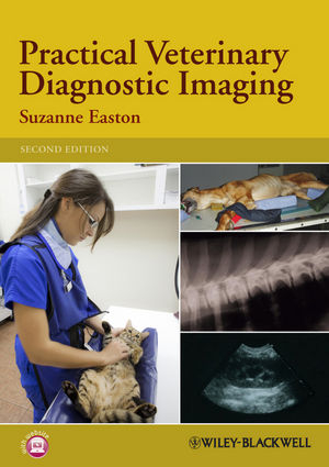
Practical Veterinary Diagnostic Imaging
Wiley-Blackwell (Verlag)
978-0-470-65648-8 (ISBN)
Practical Veterinary Diagnostic Imaging is an essential and practical guide to the various diagnostic imaging modalities that are used in veterinary practice. It moves from basic mathematic and physical principles through to discussion of equipment and practical methods. Radiographic techniques for both small and large animals are covered. There is a separate chapter devoted to ultrasound, as well as discussion of advanced imaging techniques such as fluoroscopy, computerised tomography and magnetic resonance imaging. The book also covers legislation and safety issues in the context of their impact on the veterinary practice.
Presented with clear line diagrams and photographs, Practical Veterinary Diagnostic Imaging also provides revision points and self-assessment questions in each chapter to aid study. It is an ideal guide for student and qualified veterinary nurses. It is also a useful reference for veterinary students and veterinarians in general practice who want a basic guide to radiography and other imaging modalities.
KEY FEATURES
Everything you need to know about diagnostic imaging in veterinary practice in a language you can easily understand
The basic principles of physics presented in simple terms
Improves your positioning techniques with practical tips for best practice
Clear guidance on the use of digital imaging to improve image quality and reduce radiation doses to patients
Companion website with additional resources (available at www.wiley.com/go/easton/diagnosticimaging)
Suzanne Easton is Senior Lecturer specialising in diagnostic imaging in the Faculty of Health and Life Sciences at the University of the West of England.
Figure Acknowledgements xi
1 Essential Mathematics and Physics 1
Matter, energy, power and heat 1
Units and prefixes used in radiography 3
Radiological units 4
Useful mathematics 7
Proportions and the inverse square law 7
2 The Principles of Physics Used in Radiography 11
Electrostatics – the electric charge 12
Conductors and insulators 14
Electricity 14
Measuring electricity 14
Types of current 15
Laws of an electric current 16
Resistance 16
Making a circuit – the options 17
Magnetism 17
The function and composition of a magnet 19
Magnetic laws 20
Electromagnetism – electricity and magnetism in union 21
Laws of electromagnetic induction 22
Further reading 23
3 Inside the Atom 25
Atoms, elements and other definitions 26
The ‘Make-Up’ of an atom – atomic structure 27
Shells and energy 28
The periodic table 28
Radioactivity 30
The effects of an electron changing orbits 30
Electromagnetic radiation 31
Frequency and wavelength 32
Further reading 33
4 The X-ray Tube 35
The tube housing 37
The cathode 39
The anode 42
The line focus principle 44
The anode-heel effect 45
The stator assembly 45
Tube rating 46
How to look after your X-ray tube 47
Further reading 47
5 Diagnostic Equipment 49
The X-ray circuit 50
What is seen from the outside? 51
High-voltage generators 51
Rectification 51
Mains supply switch 52
Primary circuit 52
Operating console 53
Filament circuit – control of the mA 54
High-tension circuit – provision of kV 55
Making an exposure – switches, timers and interlocks 55
Types of X-ray machines 56
Health and safety requirements 59
Power rating 59
Further reading 59
6 Production of X-rays 61
Electron production 62
Target interactions 63
X-ray emission spectrum 64
Altering the emission spectrum 65
X-ray quantity 68
X-ray quality 68
Altering exposure factors 68
Exposure charts 70
Further reading 70
7 The Effects of Radiation 71
The effect of the X-ray beam striking another atom 72
Absorption 75
Attenuation 75
The effects of ionising radiation on the body 76
Luminescence 77
Further reading 78
8 Control of the Primary Beam and Scatter 79
Light beam diaphragm 80
Factors affecting scattered radiation 81
Function of grids 81
Construction of a grid 82
Types of grid 84
Choosing a grid 85
Problems with using a grid 85
Air gap technique 86
Further reading 86
9 Radiographic Film 89
Film construction 90
Types of film 93
Formation of the latent image 94
Care and storage of films 95
Film sensitivity 96
Further reading 98
10 Intensifying Screens and Cassettes 99
The construction of intensifying screens 100
Film–screen combinations 101
Film–screen contact 104
Care of intensifying screens 104
Construction of cassettes 105
Care and use of cassettes 106
Further reading 106
11 Processing the Radiographic Film 107
The stages of processing 108
Developer 111
Fixer 112
Parts of the automatic processor 114
Replenishment 116
Silver recovery 117
The darkroom 118
Control of substances hazardous to health (COSHH) regulations 121
Other methods of processing 121
Further reading 122
12 Digital Radiography 125
Computed radiography 127
Care of the imaging plate and cassette 129
Computerised radiography process 129
Digital radiography 131
Image storage 133
Image display 134
Image quality 135
Further reading 135
13 Radiographic Image Quality 137
Sensitometry 138
Densitometry 138
Characteristic curve 139
Latitude 140
Density 141
Contrast 141
Magnification 144
Distortion 144
Movement 145
Producing a high-quality radiograph 146
Commonly seen film faults 147
Further reading 152
14 Radiation Protection 153
The effects of ionising radiation on the body 154
The basics to remember 154
Ionising Radiation Regulations 1999 155
Radiation safety in the veterinary practice 155
Classifying the areas around an X-ray machine 156
Dose limits 157
Monitoring devices 158
Lead shielding 159
Quality assurance 160
Further reading 161
15 Radiography Principles 163
General principles 164
Restraint 164
Positioning aids 165
Markers and legends 165
Assessing the radiograph 166
Terminology 166
BVA/KC hip dysplasia and elbow scoring scheme 168
Further reading 169
16 Contrast Media 171
Negative contrast medium 172
Positive contrast medium 172
Contrast examination procedures 175
Myelography 182
Other contrast examinations 184
Further reading 186
17 Small Animal Radiography Techniques 189
Chest 189
Abdomen 191
Head and neck 192
Distal extremities 196
Shoulder 198
Pelvis 200
Spine 201
Small mammals 202
Birds 203
Reptiles 204
18 Large Animal Radiography Techniques 205
Foot 205
Fetlock 207
Metacarpus and metatarsus (cannon and splint) 209
Carpus 209
Elbow 211
Shoulder 212
Tarsus 213
Stifle 214
Head 216
Spine 216
Chest 217
19 Introduction to Ultrasound 219
Sound waves 220
Ultrasound 220
How ultrasound works 220
Types of ultrasound scan 222
Doppler ultrasound 223
Effects on tissue 224
Ultrasound machines and transducers 224
Patient preparation 225
Areas suitable for examination 225
Further reading 226
20 Advance Imaging Techniques 227
Fluoroscopy 228
Computerised tomography (CT) 230
Magnetic resonance imaging (MRI) 232
Nuclear scintigraphy 234
Further reading 238
Index 239
| Erscheint lt. Verlag | 23.7.2012 |
|---|---|
| Verlagsort | Hoboken |
| Sprache | englisch |
| Maße | 175 x 246 mm |
| Gewicht | 513 g |
| Themenwelt | Veterinärmedizin ► Klinische Fächer ► Bildgebende Verfahren |
| ISBN-10 | 0-470-65648-4 / 0470656484 |
| ISBN-13 | 978-0-470-65648-8 / 9780470656488 |
| Zustand | Neuware |
| Informationen gemäß Produktsicherheitsverordnung (GPSR) | |
| Haben Sie eine Frage zum Produkt? |
aus dem Bereich


