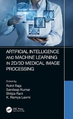
Artificial Intelligence and Machine Learning in 2D/3D Medical Image Processing
CRC Press (Verlag)
978-0-367-37435-8 (ISBN)
Digital images have several benefits, such as faster and inexpensive processing cost, easy storage and communication, immediate quality assessment, multiple copying while preserving quality, swift and economical reproduction, and adaptable manipulation. Digital medical images play a vital role in everyday life. Medical imaging is the process of producing visible images of inner structures of the body for scientific and medical study and treatment as well as a view of the function of interior tissues. This process pursues disorder identification and management.
Medical imaging in 2D and 3D includes many techniques and operations such as image gaining, storage, presentation, and communication. The 2D and 3D images can be processed in multiple dimensions. Depending on the requirement of a specific problem, one must identify various features of 2D or 3D images while applying suitable algorithms. These image processing techniques began in the 1960s and were used in such fields as space, clinical purposes, the arts, and television image improvement. In the 1970s, with the development of computer systems, the cost of image processing was reduced and processes became faster. In the 2000s, image processing became quicker, inexpensive, and simpler. In the 2020s, image processing has become a more accurate, more efficient, and self-learning technology.
This book highlights the framework of the robust and novel methods for medical image processing techniques in 2D and 3D. The chapters explore existing and emerging image challenges and opportunities in the medical field using various medical image processing techniques. The book discusses real-time applications for artificial intelligence and machine learning in medical image processing. The authors also discuss implementation strategies and future research directions for the design and application requirements of these systems.
This book will benefit researchers in the medical image processing field as well as those looking to promote the mutual understanding of researchers within different disciplines that incorporate AI and machine learning.
FEATURES
Highlights the framework of robust and novel methods for medical image processing techniques
Discusses implementation strategies and future research directions for the design and application requirements of medical imaging
Examines real-time application needs
Explores existing and emerging image challenges and opportunities in the medical field
Rohit Raja, Sandeep Kumar, Shilpa Rani, K. Ramya Laxmi
1. An Introduction to Medical Image Analysis in 3D 2. Automated Epilepsy Seizure Detection from EEG Signals Using Deep CNN Model 3. Medical Image De-Noising Using Combined Bayes Shrink and Total Variation Techniques 4. Detection of Nodule and Lung Segmentation Using Local Gabor XOR Pattern in CT Images 5. Medical Image Fusion Using Adaptive Neuro Fuzzy Inference System 6. Medical Imaging in Healthcare Applications 7. Classification of Diabetic Retinopathy by Applying an Ensemble of Architectures 8. Compression of Clinical Images Using Different Wavelet Function 9. PSO-Based Optimized Machine Learning Algorithms for the Prediction of Alzheimer’s Disease 10. Parkinson’s Disease Detection Using Voice Measurements 11. Speech Impairment Using Hybrid Model of Machine Learning 12. Advanced Ensemble Machine Learning Model for Balanced BioAssays 13. Lung Segmentation and Nodule Detection in 3D Medical Images Using Convolution Neural Network
| Erscheinungsdatum | 15.01.2021 |
|---|---|
| Zusatzinfo | 24 Tables, black and white; 105 Illustrations, black and white |
| Verlagsort | London |
| Sprache | englisch |
| Maße | 156 x 234 mm |
| Gewicht | 453 g |
| Themenwelt | Informatik ► Theorie / Studium ► Künstliche Intelligenz / Robotik |
| Medizinische Fachgebiete ► Radiologie / Bildgebende Verfahren ► Radiologie | |
| Medizin / Pharmazie ► Physiotherapie / Ergotherapie ► Orthopädie | |
| Technik ► Medizintechnik | |
| Technik ► Umwelttechnik / Biotechnologie | |
| ISBN-10 | 0-367-37435-8 / 0367374358 |
| ISBN-13 | 978-0-367-37435-8 / 9780367374358 |
| Zustand | Neuware |
| Informationen gemäß Produktsicherheitsverordnung (GPSR) | |
| Haben Sie eine Frage zum Produkt? |
aus dem Bereich


