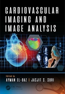
Cardiovascular Imaging and Image Analysis
Crc Press Inc (Verlag)
978-1-4987-9758-0 (ISBN)
- Titel z.Zt. nicht lieferbar
- Versandkostenfrei
- Auch auf Rechnung
- Artikel merken
This book covers the state-of-the-art approaches for automated non-invasive systems for early cardiovascular disease diagnosis. It includes several prominent imaging modalities such as MRI, CT, and PET technologies. There is a special emphasis placed on automated imaging analysis techniques, which are important to biomedical imaging analysis of the cardiovascular system. Novel 4D based approach is a unique characteristic of this product. This is a comprehensive multi-contributed reference work that will detail the latest developments in spatial, temporal, and functional cardiac imaging. The main aim of this book is to help advance scientific research within the broad field of early detection of cardiovascular disease. This book focuses on major trends and challenges in this area, and it presents work aimed to identify new techniques and their use in biomedical image analysis.
Key Features:
Includes state-of-the art 4D cardiac image analysis
Explores the aspect of automated segmentation of cardiac CT and MR images utilizing both 3D and 4D techniques
Provides a novel procedure for improving full-cardiac strain estimation in 3D image appearance characteristics
Includes extensive references at the end of each chapter to enhance further study
Ayman El-Baz is a Professor, University Scholar, and Chair of the Bioengineering Department at the University of Louisville, KY. Dr. El-Baz earned his bachelor's and master degrees in Electrical Engineering in 1997 and 2001, respectively. He earned his doctoral degree in electrical engineering from the University of Louisville in 2006. In 2009, Dr. El-Baz was named a Coulter Fellow for his contributions to the field of biomedical translational research. Dr. El-Baz has 15 years of hands-on experience in the fields of bio-imaging modeling and non-invasive computer-assisted diagnosis systems. He has authored or coauthored more than 450 technical articles (105 journals, 15 books, 50 book chapters, 175 refereed-conference papers, 100 abstracts, and 15 US patents). Jasjit S.Suri has spent over 25 years in industry as an innovator, scientist, a visionary, and is an internationally known world leader in field of Biomedical Engineering and its management. He received his Doctorate from University of Washington, Seattle, Washington, MBA from Weatherhead School of Management (WSOM), Case Western Reserve University, Cleveland, Ohio. Dr. Suri was crowned with President’s Gold medal in 1980 and the Fellow of American Institute of Medical and Biological Engineering by National Academy of Sciences in 2004 for his outstanding contributions.
A Novel 4D PDE-Based Approach for Accurate Assessment of Myocardium Function Using Cine Cardiac Magnetic Resonance Images. Magnetic Resonance Imaging Evaluation of Left Ventricular Dimentions and Function of Pericardial and Myocardial Disease. Improving Full-Cardiach Cycle Strain Estimation from Tagged CMR by Accurate Modeling of 4D & #D image Appearance Characteriistics. New Automated Markov-Gibbs Random Field Based Framework for Myocardial Wall Viability Quantification on Agent Enhanced Cardiac Magentic Resonance Images. Accurate Automatic Analysis of Cardiac Cine Images. Segmentation of the Left Ventricle from Cardiac MR Images Based on Radial GVF Snake. 4D Deformable Models with Temporal Constraints: Application to 4D Cardiac Image Segmentation. Multistage Hybrid Active Apperance Model Matching: Segmentation of Left and Right Ventricles in Cardiac MR Images. Automatic Detection of Left Ventricle in 4D MR Images. Fully Automated Framework for the Analysis of Myocardial First-Pass Perfusion MR Images. First-Pass Myocardial Perfusion Cardiovascular Magnetic Resonance at 3 Tesla. Quantitative Analysis of First-Pass Contrast-Enhanced Myocardial Perfusion MRI. Unsurpassed Inline Analysis of Cardiac Perfusion MRI. Model-Based Registration for Dynamic Cardiac Perfusion MRI.Improved Semi-Automated Segmentation of Cardiac CT and MR Images.
| Erscheinungsdatum | 17.10.2018 |
|---|---|
| Zusatzinfo | 100 Illustrations, color |
| Verlagsort | Bosa Roca |
| Sprache | englisch |
| Maße | 178 x 254 mm |
| Gewicht | 1030 g |
| Themenwelt | Medizin / Pharmazie ► Allgemeines / Lexika |
| Medizinische Fachgebiete ► Innere Medizin ► Kardiologie / Angiologie | |
| Medizin / Pharmazie ► Physiotherapie / Ergotherapie ► Orthopädie | |
| Technik ► Medizintechnik | |
| Technik ► Umwelttechnik / Biotechnologie | |
| ISBN-10 | 1-4987-9758-X / 149879758X |
| ISBN-13 | 978-1-4987-9758-0 / 9781498797580 |
| Zustand | Neuware |
| Haben Sie eine Frage zum Produkt? |
aus dem Bereich


