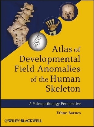
Atlas of Developmental Field Anomalies of the Human Skeleton
Wiley-Blackwell (Verlag)
978-1-118-01388-5 (ISBN)
- Titel z.Zt. nicht lieferbar
- Versandkostenfrei
- Auch auf Rechnung
- Artikel merken
Written by one of the most consulted authorities on the subject, Atlas of Developmental Field Anomalies of the Human Skeleton is the pre-eminent resource for developmental defects of the skeleton. This guide focuses on localized bone structures utilizing the morphogenetic approach that addresses the origins of variability within specific developmental fields during embryonic development. Drawings and photographs make up most of the text, forming a picture atlas with descriptive text for each group of illustrations. Each section and subdivision is accompanied by brief discussions and drawings of morphogenetic development.
Ethne Barnes is well known for her 1994 book, Developmental Defects of the Axial Skeleton, still widely in use today. A recognized specialist in the area of skeletal defects, Dr. Barnes is probably the premier expert in congenital defects of the central part of the skeleton. Currently she works as Physical Anthropologist / Paleoanthropologist for Corinth Excavations, ASCS in Tucson Arizona.
Preface xv
List of Figures xvii
INTRODUCTION 1
PART I. AXIAL SKELETON 7
A. SKULL 9
A-1. CRANIAL VAULT DEVELOPMENT 9
CRANIAL VAULT ANOMALIES 10
A-1.1. Extra Ossicles 10
A-1.2. Extra Sutures 11
A-1.3. Sutural Agenesis 11
A-1.4. Parietal Thinning 11
A-1.5. Enlarged Parietal Foramina 11
A-1.6. Inclusion Cysts 11
A-1.7. Cranial Neural Tube Defects 19
A-1.8. Hydrocephaly 20
A-1.9. Microcephaly 21
A-2. FACE DEVELOPMENT 21
FACIAL ANOMALIES 24
A-2.1. Facial Clefts 24
A-2.2. Nasal Bone Hypoplasia/Aplasia 25
A-2.3.1. Cleft Lip 25
A-2.3.2. Cleft Lip with Cleft Palate 26
A-2.4. Cleft Palate 32
A-2.5. Cleft Mandible 32
A-2.6. Mandibular Hypoplasia 32
A-2.7. Bifid Mandibular Condyle 32
A-2.8. Coronoid Hyperplasia 35
A-2.9. Palate Inclusion (Fissural) Cyst 35
A-2.10. Mandibular Inclusion Cyst 37
A-2.11. Mandibular Torus 37
A-3. EXTERNAL AUDITORY MEATUS AND TYMPANIC PLATE DEVELOPMENT 37
EXTERNAL AUDITORY MEATUS AND TYMPANIC PLATE ANOMALIES 42
A-3.1. Atresia (Aplasia)/Hypoplasia External Auditory Meatus 42
A-3.2. Tympanic Aperture 42
A-3.3. External Auditory Torus 42
A-4. STYLOHYOID CHAIN DEVELOPMENT 43
STYLOHYOID CHAIN ANOMALIES 43
A-4.1. Stylohyoid Chain Variations in Ossification 43
A-4.2. Thyroglossal Developmental Cyst 46
A-5. SKULL BASE DEVELOPMENT 49
SKULL BASE ANOMALIES 50
A-5.1. Basioccipital Hypoplasia/Aplasia 50
A-5.2. Basioccipital Clefts 50
OCCIPITAL–CERVICAL (O-C) BORDER DEVELOPMENT 50
A-5.3. Cranial Shifting of the O-C Border 50
A-5.4. Caudal Shifting of the O-C Border 55
B. VERTEBRAL COLUMN 59
VERTEBRAL COLUMN DEVELOPMENT 59
VERTEBRAL COLUMN ANOMALIES 61
B-1. Vertebral Border Shifting 61
B-1.1. Cranial Shifts of the Cervical–Thoracic (C-T) Border 61
B-1.2. Caudal Shifts of the C-T Border 61
B-1.3. Cranial Shifts of the Thoracic–Lumbar (T-L) Border 61
B-1.4. Caudal Shifts of the T-L Border 65
B-1.5. Cranial Shifts of the Lumbar–Sacral (L-S) Border 65
B-1.6. Caudal Shifts of the L-S Border 68
B-1.7. Cranial Shifts of the Sacral–Caudal (S-C) Border 70
B-1.8. Caudal Shifts of the S-C Border 70
B-2. Extra Vertebral Segment (Transitional Vertebra) 70
B-3. Cleft Neural Arch 71
B-4. Cleft Atlas Anterior Arch 74
B-5.1. Notochord Defect: Sagittal Cleft Vertebra 75
B-5.2. Notochord Defect Diastematomyelia 76
B-6. Neural Tube Defect Spina Bifi da 76
B-7. Hemivertebra: Hemimetameric Shifts 80
B-8. Lateral Hypoplasia/Aplasia 81
B-9. Ventral Hypoplasia/Aplasia 81
B-10. Dorsal Hypoplasia/Aplasia 88
B-11.1. Single Block Vertebra 92
B-11.2. Multiple Block Vertebra 92
B-11.3. Klippel–Feil Multiple Block Vertebra 93
B-12. Neural Arch Complex Disorders 93
B-13. Atlas Posterior/Lateral Bridging 95
B-14. Multiple Vertebral Anomalies 97
B-15. Sacral Agenesis versus Hemisacrum 97
B-16. Enlarged Anterior Basivertebral Foramina 103
C. RIBS 105
RIB DEVELOPMENT 105
RIB ANOMALIES 106
C-1. Supernumerary Ribs 106
Transitional Vertebra Extra Rib 106
Intrathoracic Rib 106
C-2. Rib Hypoplasia/Aplasia 106
C-3. Merged Ribs 107
C-4. Bifurcated Ribs 107
C-5. Other Rib Disorders 107
Bridged Ribs 107
Rib Spur 108
Flared Rib 108
Rib Hyperplasia 108
D. STERNUM 109
STERNUM DEVELOPMENT 109
STERNUM ANOMALIES AND VARIATIONS 109
D-1. Suprasternal Ossicles 109
D-2. Mesosternum Shape Variations 110
D-3. Manubrium–Mesosternal Joint Fusion 116
D-4. Misplaced Manubrium–Mesosternal Joint 116
D-5. Mesosternal Hypoplasia/Aplasia 117
D-6. Sternal Hyperplasia 117
D-7. Sternal Aperture 117
D-8. Sternal Caudal Clefting 118
D-9. Bifurcated Sternum 118
D-10. Pectus Excavatum (Funnel Chest) 119
D-11. Pectus Carinatum (Pigeon Breast) 120
PART II. APPENDICULAR SKELETON 121
E. UPPER LIMBS 123
UPPER LIMB DEVELOPMENT 123
SHOULDER GIRDLE SEGMENT 124
E-1. CLAVICLE DEVELOPMENT 124
CLAVICLE ANOMALIES 124
E-1.1. Clavicle Hypoplasia/Aplasia 124
E-1.2. Bifurcated Clavicle (Congenital Pseudoarthrosis) 125
E-1.3. Clavicle Duplication 125
E-2. SCAPULA DEVELOPMENT 126
SCAPULA ANOMALIES 126
E-2.1. Scapular Secondary Ossicles 126
E-2.2. Scapula Secondary Ossifi cation Hypoplasia/Aplasia 126
E-2.3. Scapula Glenoid Neck Hypoplasia 128
E-2.4. Scapular Aperture 128
E-2.5. Sprengel’s Deformity of the Scapula 128
E-2.6. Scapular Coracoid–Clavicular Bony Bridge 130
ARM SEGMENT 130
E-3. HUMERUS DEVELOPMENT 130
HUMERUS ANOMALIES 131
E-3.1. Phocomelia 131
Proximal Phocomelia (Agenesis of the Humerus) 131
Distal Phocomelia (Agenesis of the Forearm) 131
E-3.2. Proximal Humeral Head Disturbance 131
E-3.3. Distal Humerus Disturbances 131
Supracondylar Process 131
Septal Aperture 131
Nonunion of Distal Secondary Ossifications 132
Aplasia of Distal Secondary Ossifications 132
E-3.4. Elbow Patella Cubiti 132
FOREARM AND HAND SEGMENTS 132
PARAXIAL DEVELOPMENT 132
E-4. RADIUS AND ULNA DEVELOPMENT 135
RADIUS AND ULNA ANOMALIES 136
E-4.1. Forearm Meromelia (Congenital Amputation) 136
E-4.2. Forearm Paraxial Hemimelia 136
Radial (Preaxial) Hemimelia 137
Ulnar (Postaxial) Hemimelia 137
E-4.3. Duplication (Dimelia) Forearm Ray 139
E-4.4. Madelung’s Deformity 139
E-4.5. Radial–Ulnar Synostosis 139
E-4.6. Ulnar Styloid Os/Aplasia 140
E-5. CARPUS DEVELOPMENT 140
CARPAL ANOMALIES 142
E-5.1. Carpal Coalitions 142
E-5.2. Atypical Carpal Coalitions 142
Massive Carpal Coalition 144
E-5.3. Carpals Bipartite and Separated Marginal Carpal Elements 144
E-5.4. Carpal Hypoplasia/Aplasia/Hyperplasia 144
E-5.5. Os Metastyloideum 150
E-6. DIGITAL DEVELOPMENT 150
DIGITAL ANOMALIES 150
E-6.1. Brachydactyly 150
Atypical Brachydactyly 151
E-6.2. Syndactyly Complex 154
E-6.3. Symphalangism 154
E-6.4. Triphalangeal Thumb 155
E-6.5. Ectrodactyly 158
E-6.6. Polydactyly 160
F. LOWER LIMBS 163
LOWER LIMB DEVELOPMENT 163
PELVIC GIRDLE SEGMENT 164
F-1. INNOMINATE DEVELOPMENT 164
INNOMINATE ANOMALIES 165
F-1.1. Developmental Hip Dysplasia 165
F-1.2. Sacroiliac Coalition 167
THIGH SEGMENT 168
F-2. FEMUR DEVELOPMENT 168
FEMUR ANOMALIES 168
F-2.1. Proximal Femur Variations 168
Asymmetrical Torsion of the Femoral Neck 168
Hypoplasia of the Femoral Head and/or Neck 168
Coxa Vara 168
Coxa Valga 168
F-2.2. Femur Hypoplasia/Aplasia 168
Proximal Femoral Focal Defi ciency 168
Phocomelia 170
F-2.3. Bifurcated Distal Femur 170
F-3. PATELLA DEVELOPMENT 170
PATELLA ANOMALIES 170
F-3.1. Patella Hypoplasia/Aplasia 170
F-3.2. Segmented Patella 170
LOWER LEG AND FOOT SEGMENTS 171
PARAXIAL DEVELOPMENT 171
F-4. TIBIA AND FIBULA DEVELOPMENT 173
TIBIA AND FIBULA ANOMALIES 173
F-4.1. Lower Leg Meromelia (Congenital Amputation) 173
F-4.2. Lower Leg Paraxial Hemimelia 173
Tibial (Preaxial) Hemimelia 174
Fibular (Postaxial) Hemimelia 174
F-4.3. Duplication (Dimelia) Lower Leg Ray 174
F-4.4. Tibia–Fibula Synostosis 174
F-5. TARSUS DEVELOPMENT 177
TARSAL ANOMALIES 179
F-5.1. Club Foot (Talipes Equinovarus) 179
F-5.2. Vertical Talus 180
F-5.3. Tarsal Coalitions 180
F-5.4. Tarsal–Metatarsal Coalitions 182
F-5.5. Metatarsal–Phalanx Coalitions 183
F-5.6. Tibia–Hindfoot Coalition 183
F-5.7. Tarsals Bipartite and Separate Marginal Elements 183
F-5.8. Tarsal Hyperplasia/Hypoplasia/Aplasia 187
F-6. DIGITAL DEVELOPMENT 188
DIGITAL ANOMALIES 188
F-6.1. Os Metatarsium and Os Vesalianum 188
F-6.2. Brachydactyly 188
F-6.3. Syndactyly Complex 191
F-6.4. Symphalangism 191
F-6.5. Ectrodactyly 193
F-6.6. Polydactyly 193
Literature Cited 199
Index 203
| Erscheint lt. Verlag | 7.12.2012 |
|---|---|
| Verlagsort | Hoboken |
| Sprache | englisch |
| Maße | 221 x 287 mm |
| Gewicht | 885 g |
| Themenwelt | Medizin / Pharmazie ► Medizinische Fachgebiete ► Orthopädie |
| Studium ► 2. Studienabschnitt (Klinik) ► Pathologie | |
| Naturwissenschaften ► Biologie | |
| Sozialwissenschaften ► Soziologie | |
| ISBN-10 | 1-118-01388-3 / 1118013883 |
| ISBN-13 | 978-1-118-01388-5 / 9781118013885 |
| Zustand | Neuware |
| Haben Sie eine Frage zum Produkt? |
aus dem Bereich


