Clinical Nuclear Medicine Physics with MATLAB®
CRC Press (Verlag)
978-0-367-75607-9 (ISBN)
The use of MATLAB® in clinical Medical Physics is continuously increasing, thanks to new technologies and developments in the field. However, there is a lack of practical guidance for students, researchers, and medical professionals on how to incorporate it into their work.
Focusing on the areas of diagnostic Nuclear Medicine and Radiation Oncology Imaging, this book provides a comprehensive treatment of the use of MATLAB in clinical Medical Physics, in Nuclear Medicine. It is an invaluable guide for medical physicists and researchers, in addition to postgraduates in medical physics or biomedical engineering, preparing for a career in the field.
In the field of Nuclear Medicine, MATLAB enables quantitative analysis and the visualization of nuclear medical images of several modalities, such as Single Photon Emission Computed Tomography (SPECT), Positron Emission Tomography (PET), or a hybrid system where a Computed Tomography system is incorporated into a SPECT or PET system or similarly, a Magnetic Resonance Imaging system (MRI) into a SPECT or PET system. Through a high-performance interactive software, MATLAB also allows matrix computation, simulation, quantitative analysis, image processing, and algorithm implementation.
MATLAB can provide medical physicists with the necessary tools for analyzing and visualizing medical images. It is useful in creating imaging algorithms for diagnostic and therapeutic purposes, solving problems of image reconstruction, processing, and calculating absorbed doses with accuracy. An important feature of this application of MATLAB is that the results are completely reliable and are not dependent on any specific γ-cameras and workstations.
The use of MATLAB algorithms can greatly assist in the exploration of the anatomy and functions of the human body, offering accurate and precise results in Nuclear Medicine studies.
KEY FEATURES
Presents a practical, case-based approach whilst remaining accessible to students
Contains chapter contributions from subject area specialists across the field
Includes real clinical problems and examples, with worked through solutions
Maria Lyra Georgosopoulou, PhD, is a Medical Physicist and Associate Professor at the National and Kapodistrian University of Athens, Greece.
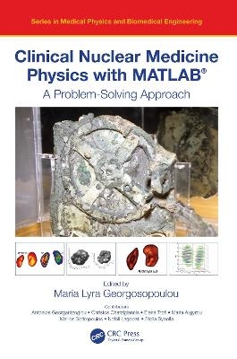
Photo credit: The Antikythera Mechanism is the world’s oldest known analog computer. It consisted of many wheels and discs that could be placed onto the mechanism for calculations. It is possible that the first algorithms and analog calculations in mathematics were implemented with this mechanism, invented in the early first centuries BC. It has been selected for the cover to demonstrate the importance of calculations in science.
Maria Lyra Georgosopoulou, PhD, is an Associate Professor and Medical Physicist. She was an Associate Professor in the National and Kapodistrian University of Athens and a Radiation Protection Officer at Aretaeion Hospital. For twenty years she was the Scientific Director of the "Paediatric Nuclear Medical Imaging Center" (IATRIKI APEIKONISI /IA) in Athens, Greece. The integrating theme of her research has been applications of advanced technology to practical biomedical problems in Nuclear Medicine and Medical Ultrasound Image Processing and Evaluation.
1. Introduction. 2. Image formation in Nuclear Medicine. 3. Nuclear Medicine Imaging Essentials. 4. Methods of Imaging Reconstruction in Nuclear Medicine. 5. Image processing and analysis in Nuclear Medicine. 6. 3D Volume data in Nuclear Medicine. 7. Quantification in Nuclear Medicine Imaging. 8. Quality Control of Nuclear Medicine Equipment. 9. Introduction to MATLAB and Basic MATLAB processes for Nuclear Medicine. 10. Morphology of human organs in Nuclear Medicine – MATLAB commands. 11. Internal Dosimetry by MATLAB in Therapeutic Nuclear Medicine. 12. Pharmacokinetics in Nuclear Medicine-MATLAB use. 13. Nanotechnology in Nuclear Medicine-MATLAB use. 14. CASE Studies in Nuclear Medicine/ MATLAB Approach.
| Erscheinungsdatum | 24.08.2021 |
|---|---|
| Reihe/Serie | Series in Medical Physics and Biomedical Engineering |
| Zusatzinfo | 13 Tables, black and white; 32 Line drawings, color; 25 Line drawings, black and white; 32 Halftones, color; 26 Halftones, black and white; 64 Illustrations, color; 51 Illustrations, black and white |
| Verlagsort | London |
| Sprache | englisch |
| Maße | 156 x 234 mm |
| Gewicht | 453 g |
| Themenwelt | Sachbuch/Ratgeber ► Gesundheit / Leben / Psychologie |
| Medizin / Pharmazie ► Allgemeines / Lexika | |
| Medizinische Fachgebiete ► Radiologie / Bildgebende Verfahren ► Nuklearmedizin | |
| Medizinische Fachgebiete ► Radiologie / Bildgebende Verfahren ► Radiologie | |
| Naturwissenschaften ► Biologie | |
| Naturwissenschaften ► Physik / Astronomie ► Atom- / Kern- / Molekularphysik | |
| Technik | |
| ISBN-10 | 0-367-75607-2 / 0367756072 |
| ISBN-13 | 978-0-367-75607-9 / 9780367756079 |
| Zustand | Neuware |
| Informationen gemäß Produktsicherheitsverordnung (GPSR) | |
| Haben Sie eine Frage zum Produkt? |
aus dem Bereich


