Human Brain Anatomy in Computerized Images
Oxford University Press Inc (Verlag)
978-0-19-516561-6 (ISBN)
- Titel ist leider vergriffen;
keine Neuauflage - Artikel merken
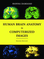
Modern tomographic scans are revealing the structure of the human brain in unprecedented detail. This spectator progress, however, poses a critical problem for neuroscientists and practitioners of brain-related professions: how to find their way in the current tomographic images so as to identify a particular brain site, be it normal or damaged by disease? The problem is made all the more difficult by the large degree of individual neuroanatomical variation.
Prepared by a leading expert in advanced brain-imaging techniques, this unique atlas is a guide to the localization of brain structures that illustrates the wide range of neuranatomical variation. It is based on the analysis of 29 normal brain obtained from three-dimensional reconstructions of magnetic
resonance scans of living persons. It also provides 177 sections (coronal, axial, and parasagital) of one of those brains so that the same structure presented in the section obtained in one incidence can be identified in the section of another incidence. An additional 209 sections of two incidences of two other brains with different overall configurations are included at the same incidences, so that readers can become familiar with the variability of standard images prompted by different
skull shapes. Forty-six normal brains, segmented in to the major lobes, are also included. The atlas is based on a voxel-rendering technique developed in the author's laboratory that permits the reconstruction of the brain in three dimensions. The technique permits the identification of major sulci
and gyri with about the same degree of precision that can be achieved at the autopsy table. The volume contains 50 pages of colour illustrations. The Second Edition of this atlas offers entirely new images, all from new brain specimens. Like the first edition, it will prove to be an essential tool for neurologists, neurosurgeons, neuroradiologists, psychiatrists, and neuroscientists, as well as medical and neuroscience students.
Hanna Damasio is Distinguished Professor of Neurology at The University of Iowa College of Medicine, where she directs the Human Neuroanatomy and Neuroimaging Laboratory, and Adjunct Professor at the Salk Institute. Using computerized tomography and magnetic resonance scanning, she developed methods of investigating human brain structure and studied functions such as language using both the lesion method and functional neuroimaging. She is the author of over 150 scientific publications and of the award-winning Lesion Analysis in Neuropsychology (Oxford University Press), which has been used worldwide in brain-imaging work. Damasio is a Fellow of the American Academy of Arts and Sciences and of the American Neurological Association. She recently shared the Signoret Prize in cognitive neurosciences with Antonio Damasio. She holds honorary doctorates from the Universities of Lisbon and Aachen. Since 1992 she has been listed in Best Doctors in America under Neurology.
REFERENCES
| Erscheint lt. Verlag | 12.5.2005 |
|---|---|
| Zusatzinfo | halftones and colour figures throughout |
| Verlagsort | New York |
| Sprache | englisch |
| Maße | 237 x 313 mm |
| Gewicht | 2430 g |
| Themenwelt | Medizin / Pharmazie ► Medizinische Fachgebiete ► Neurologie |
| Medizin / Pharmazie ► Medizinische Fachgebiete ► Radiologie / Bildgebende Verfahren | |
| Naturwissenschaften ► Biologie ► Humanbiologie | |
| Naturwissenschaften ► Biologie ► Zoologie | |
| ISBN-10 | 0-19-516561-6 / 0195165616 |
| ISBN-13 | 978-0-19-516561-6 / 9780195165616 |
| Zustand | Neuware |
| Haben Sie eine Frage zum Produkt? |
aus dem Bereich


