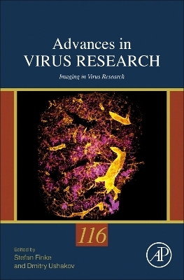
Imaging in Virus Research
Academic Press Inc (Verlag)
978-0-443-15814-8 (ISBN)
Prof. Dr. Stefan Finke, is the Head of the Institute and Laboratory Head of the Institute of Molecular Virology and Cell Biology (IMVZ) of Friedrich-Loeffler-Institut, the Federal Research Institute for Animal Health of Germany. Dmitry Ushakov is a Head of Imaging and Bioinformatics laboratory at Friedrich Loeffler Institute, Riems Island. His current research is centered on understanding the fundamental principles governing virus-host interactions and associated immune cell responses. He obtained his PhD in Biochemistry at the University of North Texas Health Science Center at Fort Worth. In his early research carrier at Imperial College London, Hanover Medical School and University of Leeds, he carried out development of advanced fluorescence imaging methods with modalities ranging from high-resolution imaging, to ratiometric imaging, such as fluorescence lifetime imaging microscopy, to super-resolution fluorescence microscopy. He applied them to studies across scales investigating biological motility, organelle traffic and signalling in immune, epithelial and muscle cells. He later became interested in cell behavior in complex 3D environment of biological tissues in homeostasis and in response to stress or infection. Working at King’s College London and the Crick Institute, he developed a high-throughput immune cell screening platform, which combined confocal microscopy with automated multi-parameter quantitative 3D image analysis. Recently his laboratory established novel approaches for tissue optical clearing, volumetric imaging and 3D image analysis enabling quantification of large tissue volumes and whole organs in different animals. This technology has been applied to study infection by a number of neurotropic and respiratory viruses, such as Rabies virus and SARS-CoV-2.
1. Recent developments of advanced fluorescence microscopy methods for biological applications 2. Fluorescence microscopy to image virus entry: Probing different angles Mario Schelhaas 3. RSV and cytoplasmic inclusion bodies Jennifer Risso Ballester and Marie-Anne Rameix-Welti 4. Advanced Imaging of HIV-1 fusion and virus-host lipid interactions Harshitha Ramu and Sergi Padilla-Parra 5. Spatiotemporal orchestration of virus morphogenesis Jens Bosse 6. Imaging applied to study interactions between poxviruses and their host cells Jason P. Mercer 7. Viral capsid structures Yuliia Mironova and Kay Grünewald 8. Tissue Clearing and 3D-Imaging of Virus Infections Dmitry Ushakov and Stefan Finke 9. High-resolution fluorescence microscopy applied for airborne virus imaging Christian Sieben 10. Multimode imaging of phase separation and RNA virus factory morphogenesis Roma Tuma
| Erscheinungsdatum | 03.08.2023 |
|---|---|
| Reihe/Serie | Advances in Virus Research |
| Verlagsort | San Diego |
| Sprache | englisch |
| Maße | 152 x 229 mm |
| Gewicht | 480 g |
| Themenwelt | Medizin / Pharmazie ► Medizinische Fachgebiete ► Mikrobiologie / Infektologie / Reisemedizin |
| Studium ► Querschnittsbereiche ► Infektiologie / Immunologie | |
| Studium ► Querschnittsbereiche ► Prävention / Gesundheitsförderung | |
| Naturwissenschaften ► Biologie ► Mikrobiologie / Immunologie | |
| ISBN-10 | 0-443-15814-2 / 0443158142 |
| ISBN-13 | 978-0-443-15814-8 / 9780443158148 |
| Zustand | Neuware |
| Informationen gemäß Produktsicherheitsverordnung (GPSR) | |
| Haben Sie eine Frage zum Produkt? |
aus dem Bereich


