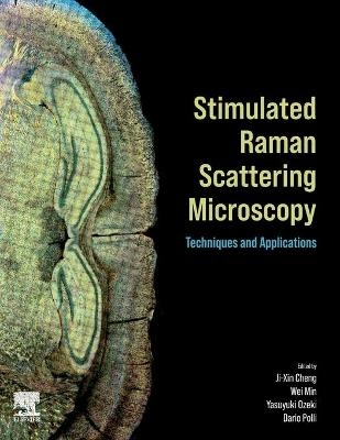
Stimulated Raman Scattering Microscopy
Elsevier - Health Sciences Division (Verlag)
978-0-323-85158-9 (ISBN)
This rapidly growing field needs a comprehensive resource that brings together the current knowledge on the topic, and this book does just that. Researchers who need to know the requirements for all aspects of the instrumentation as well as the requirements of different imaging applications (such as different types of biological tissue) will benefit enormously from the examples of successful demonstrations of SRS imaging in the book.
Led by Editor-in-Chief Ji-Xin Cheng, a pioneer in coherent Raman scattering microscopy, the editorial team has brought together various experts on each aspect of SRS imaging from around the world to provide an authoritative guide to this increasingly important imaging technique. This book is a comprehensive reference for researchers, faculty, postdoctoral researchers, and engineers.
Dr. Ji-Xin Cheng attended University of Science and Technology of China (USTC) from 1989 to 1994. He carried out his PhD study on bond-selective chemistry at USTC. As a graduate student, he worked as a research assistant at Universite Paris-sud on vibrational spectroscopy and the Hong Kong University of Science and Technology (HKUST) on quantum dynamics theory. After postdoctoral training on ultrafast spectroscopy at HKUST, he joined Sunney Xie’s group at Harvard University as a postdoc and worked on the development of CARS microscopy. Cheng joined Purdue University as Assistant Professor in 2003, promoted to Associate Professor in 2009 and Full Professor in 2013. He joined Boston University as the Inaugural Theodore Moustakas Chair Professor in Photonics and Optoelectronics in 2017. For his pioneering contributions to the chemical imaging field, Cheng received the 2020 Pittsburg Spectroscopy Award, the 2019 Ellis R. Lippincott Award, and the 2015 Craver Award. Dr. Wei Min graduated from Peking University in 2003. He received his Ph.D. from Harvard University in 2008 studying single-molecule biophysics with Prof. Sunney Xie. After continuing his postdoctoral work in the Xie group, Dr. Min joined the faculty at Columbia University in 2010 and was promoted to Full Professor there in 2017. Dr. Min’s current research interests focus on developing novel optical spectroscopy and microscopy technology to address biomedical problems. His group has made important contributions to the development of stimulated Raman scattering (SRS) microscopy and its broad application in biomedical imaging. Dr. Min’s contribution has been recognized by a number of honors, including Scientific Achievement Award from Royal Microscopical Society (2021), Pittsburgh Conference Achievement Award (2019), Coblentz Award of Molecular Spectroscopy (2017), ACS Early Career Award in Experimental Physical Chemistry (2017), and NIH Director’s New Innovator Award (2012). Dr. Yasuyuki Ozeki received B.S., M.S. and Dr. Eng. Degrees in Electronic Engineering from the University of Tokyo, Tokyo, Japan, in 1999, 2001, and 2004, respectively. In 2004, he joined Furukawa Electric Co., Ltd., as a postdoctoral researcher of Japan Science and Technology Agency (JST). In 2006, he joined Department of Material and Life Science, Osaka University, Osaka, Japan, as an assistant professor. From 2009 to 2013, he was also PRESTO researcher of JST. In 2013, he was appointed as an Associate Professor of Department of Electrical Engineering and Information Systems, the University of Tokyo, Tokyo, Japan, and was promoted to a Full Professor in 2021. His work covers millimeter-wave photonics, nonlinear fiber optics, ultrafast lasers, and their application to microprocessing and biomedical microscopy. His current research focuses on biomedical imaging by stimulated Raman scattering (SRS) microscopy, and its related technologies including highly functional ultrafast laser sources, detection electronics, image processing, etc. Dario Polli is Associate Professor of Physics at Politecnico di Milano (Italy) since 2014, where he is heading a research group of more than 10 people including post-docs, Ph.D. and diploma students. He is affiliated with the Center for Nano Science and Technology of the Italian Institute of Technology in Milan, Italy. His main research focus is on coherent Raman spectroscopy and microscopy, ultrafast and non-linear optics, Fourier-transform spectroscopy and time-resolved pump-probe spectroscopy and microscopy. He is the recipient of many research grants, including an ERC Consolidator grant on the development of high-speed broadband coherent Raman microscopy for fast and reliable tumour identification. He also devotes to technology transfer: he filed several patents and has founded two start-up companies in the field of photonics. Finally, he is passionate about Science divulgation to the public.
Preface
Sunney Xie
Part 1: Theory
1. Coherent Raman scattering processes
2. Sensitivity and noise in SRS microscopy
3. Stimulated Raman Scattering: ensembles to single molecules
Part 2: Advanced Instrumentation and Emerging Modalities
4. Hyperspectral SRS imaging via spectral focusing
5. Balanced detection SRS microscopy
6. Multiplex stimulated Raman Scattering microscopy via a tuned amplifier
7. Impulsive SRS microscopy
8. Multicolor SRS imaging with wavelength-tunable/switchable lasers
9. Pulse-shaping-based SRS spectral imaging and applications
10. Background-free stimulated Raman scattering imaging by manipulating photons in the spectral domain
11. Coherent Raman scattering microscopy for superresolution vibrational imaging: Principles, techniques, and implementations
12. Quantum-enhanced stimulated Raman scattering
13. Stimulated Raman excited fluorescence (SREF) microscopy: Combining the best of two worlds
14. Instrumentation and methodology for volumetric stimulated Raman scattering imaging
15. SRS flow and image cytometry
16. Widely and rapidly tunable fiber laser for high-speed multicolor SRS
17. Compact fiber lasers for stimulated Raman scattering microscopy
18. Synchronized time-lens source for coherent Raman scattering microscopy
Part 3: Vibrational Probes
19. Spontaneous Raman and SERS microscopy for Raman tag imaging
20. Stimulated Raman scattering imaging with small vibrational probes
21. Supermultiplexed vibrational imaging: From probe development to biomedical application
22. Raman beads for bio-imaging
23. Plasmon-enhanced stimulated Raman scattering microscopy
Part 4: Data Science
24. Converting hyperspectral SRS into chemical maps
25. Compressive Raman microspectroscopy
26. Denoise SRS images
Part 5: Applications to Life Sciences and Materials Science
27. Use of SRS microscopy for imaging drugs
28. Isotope-probed SRS (ip-SRS) imaging of metabolic dynamics in living organisms
29. Rapid determination of antimicrobial susceptibility by SRS single-cell metabolic imaging
30. Stimulated Raman scattering imaging of cancer metabolism: New avenue to precision medicine
31. Biomedical applications of SRS microscopy in functional genetics and genomics
32. Stimulated Raman voltage imaging for quantitative mapping of membrane potential
33. Neurodegenerative disease by SRS microscopy
34. Applications of stimulated Raman scattering (SRS) microscopy in materials science
35. Resolving molecular orientation by polarization-sensitive stimulated Raman scattering microscopy
Part 6: Miniaturization and Translation to Medicine
36. Stimulated Raman histology
37. Miniaturized handheld stimulated Raman scattering microscope
38. Intraoperative multimodal imaging
| Erscheinungsdatum | 17.12.2021 |
|---|---|
| Zusatzinfo | Approx. 300 illustrations; Illustrations |
| Verlagsort | Philadelphia |
| Sprache | englisch |
| Maße | 216 x 276 mm |
| Gewicht | 1650 g |
| Themenwelt | Naturwissenschaften ► Chemie ► Analytische Chemie |
| ISBN-10 | 0-323-85158-4 / 0323851584 |
| ISBN-13 | 978-0-323-85158-9 / 9780323851589 |
| Zustand | Neuware |
| Informationen gemäß Produktsicherheitsverordnung (GPSR) | |
| Haben Sie eine Frage zum Produkt? |
aus dem Bereich


