Biomedical Visualisation
Springer International Publishing (Verlag)
978-3-030-31906-9 (ISBN)
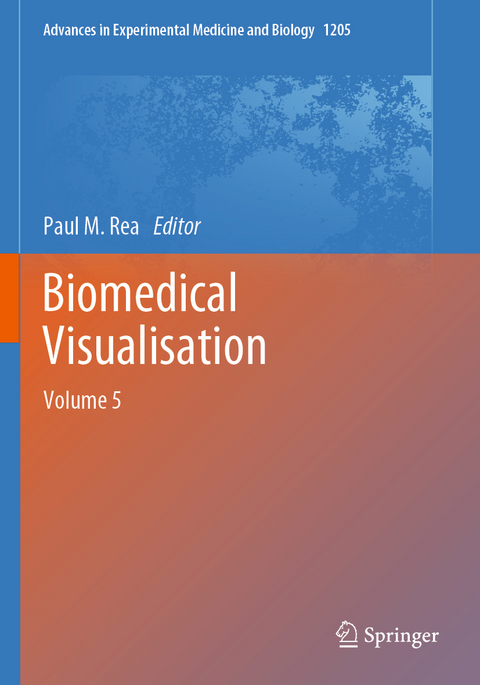
This edited volume explores the use of technology to enable us to visualise the life sciences in a more meaningful and engaging way. It will enable those interested in visualisation techniques to gain a better understanding of the applications that can be used in visualisation, imaging and analysis, education, engagement and training.
The reader will be able to explore the utilisation of technologies from a number of fields to enable an engaging and meaningful visual representation of the biomedical sciences, with a focus in this volume related to anatomy, and clinically applied scenarios.
The first four chapters highlight the diverse uses of CT and MRI scanning. These chapters demonstrate the uses of modern scanning techniques currently in use both clinically and in research and include vascular modelling, uses of the stereoscopic model, MRI in neurovascular and neurodegenerative diseases, and how they can also be used in a forensic setting in identification.The remaining six chapters truly demonstrate the diversity technology has in education, training and patient engagement. Multimodal technologies are discussed and include art and history collections, photogrammetry and games engines, augmented reality and review of the current literature for patient rehabilitation and education of the health professions. These chapters really do provide "something for everyone" whether you are a student, faculty member, or part of our curious global population interested in technology and healthcare.
lt;p>Paul is a Professor of Digital and Anatomical Education at the University of Glasgow. He is qualified with a medical degree (MBChB), a MSc (by research) in craniofacial anatomy/surgery, a PhD in neuroscience, the Diploma in Forensic Medical Science (DipFMS), and an MEd with Merit (Learning and Teaching in Higher Education). He is an elected Fellow of the Royal Society for the encouragement of Arts, Manufactures and Commerce (FRSA), elected Fellow of the Royal Society of Biology (FRSB), Senior Fellow of the Higher Education Academy, professional member of the Institute of Medical Illustrators (MIMI) and a registered medical illustrator with the Academy for Healthcare Science.
Paul has published widely and presented at many national and international meetings, including invited talks. He sits on the Executive Editorial Committee for the Journal of Visual Communication in Medicine, is Associate Editor for the European Journal of Anatomy and reviews for 24 different journals/publishers.
He is the Public Engagement and Outreach lead for anatomy coordinating collaborative projects with the Glasgow Science Centre, NHS and Royal College of Physicians and Surgeons of Glasgow. Paul is also a STEM ambassador and has visited numerous schools to undertake outreach work.
His research involves a long-standing strategic partnership with the School of Simulation and Visualisation The Glasgow School of Art. This has led to multi-million pound investment in creating world leading 3D digital datasets to be used in undergraduate and postgraduate teaching to enhance learning and assessment. This successful collaboration resulted in the creation of the worlds first taught MSc Medical Visualisation and Human Anatomy combining anatomy and digital technologies. The Institute of Medical Illustrators also accredits it. This degree, now into its 8th year, has graduated almost 100 people, and created college-wide, industry, multi-institutional and NHS research linked projects for students. Paul is the Programme Director for this degree.
Comparison of Magnetic Resonance Angiography and Computed Tomography Angiography Stereoscopic Cerebral Vascular Models.- Use Stereoscopic Model in Interventional and Surgical Procedures.- The Whole Picture: From Isolated to Global MRI Measures of Neurovascular and Neurodegenerative Disease.- Three-dimensional geometry of phalanges as a proxy for pair-matching: mesh comparison using an ICP algorithm.- Multimodal learning in health sciences and medicine: Merging technologies to enhance student learning and communication.- Dissecting Art: Developing Object-Based Teaching Using Historical Collections.- Using Photogrammetry to Create a Realistic 3D Anatomy Learning Aid with Unity Game Engine.- Innovative education and engagement tools for Rheumatology and Immunology Public Engagement with Augmented Reality.- A Game Changer: 'The Use of Digital Technologies in the Management of Upper Limb Rehabilitation'.- The Use of Social Media in Anatomicaland Health Professional Education: A Systematic Review.
| Erscheinungsdatum | 16.01.2021 |
|---|---|
| Reihe/Serie | Advances in Experimental Medicine and Biology |
| Zusatzinfo | XV, 170 p. 59 illus., 54 illus. in color. |
| Verlagsort | Cham |
| Sprache | englisch |
| Maße | 178 x 254 mm |
| Gewicht | 410 g |
| Themenwelt | Informatik ► Weitere Themen ► Bioinformatik |
| Medizin / Pharmazie ► Physiotherapie / Ergotherapie ► Orthopädie | |
| Naturwissenschaften ► Biologie ► Genetik / Molekularbiologie | |
| Technik ► Umwelttechnik / Biotechnologie | |
| Schlagworte | 3D Printing • Computing Technology • E-Tutorial • Photogrammetry • Virtual and Augmented Reality |
| ISBN-10 | 3-030-31906-7 / 3030319067 |
| ISBN-13 | 978-3-030-31906-9 / 9783030319069 |
| Zustand | Neuware |
| Informationen gemäß Produktsicherheitsverordnung (GPSR) | |
| Haben Sie eine Frage zum Produkt? |
aus dem Bereich


