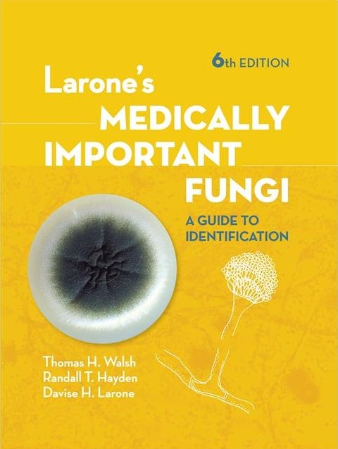
Larone's Medically Important Fungi
American Society for Microbiology (Verlag)
978-1-55581-987-3 (ISBN)
- Titel ist leider vergriffen;
keine Neuauflage - Artikel merken
Davise H. Larone is well known as the originator of the book that many readers have come to rely upon for assistance in the accurate identification of fungi from patient specimens, a key step in treating mycotic infections. Dr. Larone has now been joined by Thomas J. Walsh and Randall T. Hayden to update this gold standard reference while retaining the format that has made this guide so popular for more than 40 years.
Led by editors Siddhartha Thakur and Kalmia Kniel, a team of expert authors provides insights into critical themes surrounding preharvest food safety, including:
Medically Important Fungi will expand your knowledge and support your work by:
- Providing detailed descriptions of the major mycoses as viewed in patients’ specimens by direct microscopic examination of stained slides
- Offering a logical step-by-step process for identification of cultured organisms, utilizing detailed descriptions, images, pointers on organisms’ similarities and distinctions, and selected references for further information
- Covering nearly 150 of the fungi most commonly encountered in the clinical mycology laboratory
- Presenting details on each organism’s pathogenicity, growth characteristics, relevant biochemical reactions, and microscopic morphology, illustrated with photomicrographs, Dr. Larone's unique and elegant drawings, and color photos of colony morphology and various test results
- Explaining the current changes in fungal taxonomy and nomenclature that are due to information acquired through molecular taxonomic studies of evolutionary fungal relationships
- Providing basic information on molecular diagnostic methods, e.g., PCR amplification, nucleic acid sequencing, MALDI-TOF mass spectrometry, and other commercial platforms
- Including an extensive section of easy-to-follow lab protocols, a comprehensive list of media and stain procedures, guidance on collection and preparation of patient specimens, and an illustrated glossary
With Larone’s Medically Important Fungi: A Guide to Identification, both novices and experienced professionals in clinical microbiology laboratories can continue to confidently identify commonly encountered fungi.
Thomas J. Walsh, MD, PhD (hon), FIDSA, FAAM, FECMM, serves as Professor of Medicine, Pediatrics, and Microbiology & Immunology at Weill Cornell Medicine of Cornell University; founding Director of the Transplantation-Oncology Infectious Diseases Program and the Infectious Diseases Translational Research Laboratory, Henry Schueler Foundation Scholar in Mucormycosis; Investigator of Emerging Infectious Diseases of the Save Our Sick Kids Foundation; and Attending Physician at the NewYork Presbyterian Hospital and Hospital for Special Surgery. Dr. Walsh directs a combined clinical and laboratory research program dedicated to improving the lives and care of immunocompromised children and adults with invasive mycoses and other life-threatening infections. The objective of the Program’s translational research is to develop new strategies for laboratory diagnosis, treatment, and prevention of life-threatening invasive mycoses in immunocompromised patients. Dr. Walsh brings to this book more than three decades of experience in the field of medical mycology, with clinical and laboratory expertise across a wide spectrum of medically important fungi and mycoses. In addition to patient care and translational research, Dr. Walsh has also mentored more than 180 students, fellows, and faculty, many of whom are distinguished leaders in the field of medical mycology throughout the world.
Randall T. Hayden, MD, is Director of Clinical Pathology Laboratories and Director of Clinical and Molecular Microbiology and Member in the Department of Pathology at St. Jude Children’s Research Hospital in Memphis, Tennessee. He joined the faculty there in 2000, following postdoctoral training in microbiology and molecular microbiology at the Mayo Clinic and in surgical pathology at the MD Anderson Cancer Center. He is board certified in Anatomic and Clinical Pathology with sub-specialty certification in Medical Microbiology. His research interests focus on the application of molecular methods to diagnostic challenges in clinical microbiology, with particular emphasis on the diagnosis of infections in the immunocompromised host. He is editor-in-chief of Diagnostic Microbiology of the Immunocompromised Host, 2nd Edition; co-editor of Molecular Microbiology, Diagnostic Principles and Practice, 3rd Edition; and section editor for the Manual of Clinical Microbiology, 12th Edition, all from ASM Press.
List of Tables xv
Preface to the Fifth Edition xvii
Preface to the First Edition xix
Acknowledgments xxi
How to Use the Guide ... 1
Use of Reference Laboratories ... 3
Safety Precautions ... 7
Part I Direct Microscopic Examination of Clinical Specimens
Introduction ... 11
Histological Terminology ... 13
Tissue Reactions to Fungal Infection ... 17
Stains ... 21
Table 1 Stains for direct microscopic observation of fungi and/or filamentous bacteria in tissue ... 22
Guide to Interpretation of Direct Microscopic Examination ... 23
Detailed Descriptions ... 31
Actinomycosis ... 33
Mycetoma, Actinomycotic or Eumycotic ... 34
Nocardiosis ... 36
Zygomycosis ... 37
Aspergillosis ... 38
Miscellaneous Hyalohyphomycoses ... 40
Dermatophytosis ... 42
Tinea versicolor ... 43
Tinea nigra ... 44
Phaeohyphomycosis ... 45
Chromoblastomycosis ... 46
Sporotrichosis ... 47
Histoplasmosis capsulati ... 48
Penicilliosis marneffei ... 50
Blastomycosis ... 52
Paracoccidioidomycosis ... 53
Candidiasis (Candidosis) ... 54
Trichosporonosis ... 56
Cryptococcosis ... 57
Pneumocystosis ... 59
Protothecosis ... 60
Coccidioidomycosis ... 61
Rhinosporidiosis ... 62
Adiaspiromycosis ... 64
Special References ... 65
Part II Identification of Fungi in Culture
Guide to Identification of Fungi in Culture ... 69
Detailed Descriptions ... 101
Filamentous Bacteria ... 103
Introduction ... 105
Table 2 Differentiation of filamentous aerobic actinomycetes encountered in clinical specimens ... 107
Nocardia spp. ... 108
Table 3 Phenotypic characteristics of most common clinically encountered Nocardia spp. ... 110
Streptomyces spp. ... 111
Actinomadura spp. ... 112
Nocardiopsis dassonvillei ... 113
Yeasts and Yeastlike Organisms ... 115
Introduction ... 117
Candida albican ... 119
Table 4 Characteristics of the genera of clinically encountered yeasts and yeastlike organisms ... 120
Candida dubliniensis ... 121
Table 5 Characteristics of Candida spp. most commonly encountered in the clinical laboratory ... 122
Table 6 Characteristics that assist in differentiating Candida dubliniensis from Candida albicans ... 124
Candida tropicalis ... 125
Candida parapsilosis complex ... 126
Candida lusitaniae ... 127
Candida krusei ... 128
Table 7 Differentiating characteristics of Blastoschizomyces capitatus versus Candida krusei ... 129
Table 8 Differentiating characteristics of C. krusei, C. inconspicua, and C. norvegensis ... 129
Candida kefyr (formerly Candida pseudotropicalis) ... 130
Candida rugosa ... 131
Candida guilliermondii complex ... 132
Table 9 Differentiating characteristics of Candida guilliermondii versus Candida famata ... 133
Candida lipolytica ... 134
Candida zeylanoides ... 135
Candida glabrata ... 136
Cryptococcus neoformans ... 137
Cryptococcus gattii ... 138
Table 10 Characteristics of Cryptococcus spp. ... 139
Table 11 Characteristics of yeasts and yeastlike organisms other than Candida spp. and Cryptococcus spp. ... 140
Rhodotorula spp. ... 141
Sporobolomyces salmonicolor ... 142
Saccharomyces cerevisiae ... 143
Wickerhamomyces anomalus (formerly Pichia anomala and Hansenula anomala) (sexual state); Candida pelliculosa (asexual state) ... 145
Malassezia spp. ... 146
Malassezia pachydermatis ... 148
Ustilago sp. ... 149
Prototheca spp. ... 150
Trichosporon spp. ... 151
Table 12 Key characteristics of the most common clinically encountered Trichosporon spp. ... 152
Blastoschizomyces capitatus ... 153
Geotrichum candidum ... 154
Thermally Dimorphic Fungi ... 155
Introduction ... 157
Histoplasma capsulatum ... 158
Blastomyces dermatitidis ... 160
Paracoccidioides brasiliensis ... 162
Penicillium marneffei ... 164
Sporothrix schenckii complex ... 166
Table 13 Characteristics for differentiating species of the Sporothrix schenckii complex ... 168
Thermally Monomorphic Moulds ... 169
Zygomycetes ... 171
Introduction ... 173
Table 14 Differential characteristics of similar organisms in the class Zygomycetes ... 175
Table 15 Differential characteristics of the clinically encountered Rhizopus spp. ... 175
Rhizopus spp. ... 176
Mucor spp. ... 177
Rhizomucor spp. ... 178
Lichtheimia corymbifera complex (formerly Absidia corymbifera) ... 179
Apophysomyces elegans ... 181
Saksenaea vasiformis ... 183
Cokeromyces recurvatus ... 184
Cunninghamella bertholletiae ... 185
Syncephalastrum racemosum ... 186
Basidiobolus sp. ... 187
Conidiobolus coronatus ... 188
Dematiaceous Fungi ... 189
Introduction ... 191
Fonsecaea pedrosoi ... 193
Fonsecaea compacta ... 195
Rhinocladiella spp. ... 196
Phialophora verrucosa ... 197
Table 16 Characteristics of Phialophora, Pleurostomophora, Phaeoacremonium, Acremonium, Phialemonium, and Lecythophora ... 198
Pleurostomophora richardsiae (formerly Phialophora richardsiae) ... 199
Phaeoacremonium parasiticum (formerly Phialophora parasitica) ... 200
Phialemonium spp. ... 201
Cladosporium spp. ... 203
Table 17 Characteristics of Cladosporium and Cladophialophora spp. ... 204
Cladophialophora carrionii ... 205
Cladophialophora bantiana ... 206
Cladophialophora boppii (formerly Taeniolella boppii) ... 207
Pseudallescheria boydii (sexual state) / Scedosporium apiospermum (asexual state) complex ... 208
Table 18 Differentiating phenotypic characteristics of the clinically encountered members of the Pseudallescheria boydii complex and Scedosporium prolificans ... 210
Scedosporium prolificans (formerly Scedosporium inflatum) ... 211
Ochroconis gallopava (formerly Dactylaria constricta var.gallopava) ... 212
Table 19 Differentiation of the clinically encountered Ochroconis species ... 213
Table 20 Characteristics of some of the "black yeasts" ... 213
Exophiala jeanselmei complex ... 214
Exophiala dermatitidis (Wangiella dermatitidis) ... 215
Hortaea werneckii (Phaeoannellomyces werneckii) ... 216
Madurella mycetomatis ... 217
Madurella grisea ... 218
Piedraia hortae ... 219
Aureobasidium pullulans ... 220
Table 21 Differential characteristics of Aureobasidium pullulans versus Hormonema dematioides ... 222
Hormonema dematioides ... 223
Neoscytalidium dimidiatum (formerly Scytalidium dimidiatum) ... 224
Botrytis sp. ... 226
Stachybotrys chartarum (S. alternans, S. atra) ... 227
Graphium eumorphum ... 228
Curvularia spp. ... 229
Bipolaris spp. ... 230
Table 22 Characteristics of Bipolaris, Drechslera, and Exserohilum spp. ... 231
Exserohilum spp. ... 232
Helminthosporium sp ... 233
Alternaria sp ... 234
Ulocladium sp. ... 235
Stemphylium sp. ... 236
Pithomyces sp. ... 237
Epicoccum sp. ... 238
Nigrospora sp. ... 239
Chaetomium sp. ... 240
Phoma spp. ... 241
Dermatophytes ... 243
Introduction ... 245
Microsporum audouinii ... 247
Microsporum canis var. canis ... 248
Microsporum canis var. distortum ... 249
Microsporum cookei ... 250
Microsporum gypseum complex ... 251
Microsporum gallinae ... 252
Microsporum nanum ... 253
Microsporum vanbreuseghemii ... 254
Microsporum ferrugineum ... 255
Trichophyton mentagrophytes ... 256
Table 23 Differentiation of similar conidia-producing Trichophyton spp ... 257
Trichophyton rubrum ... 258
Trichophyton tonsurans ... 259
Trichophyton terrestre ... 260
Trichophyton megninii ... 261
Trichophyton soudanense ... 262
Table 24 Growth patterns of Trichophyton species on nutritional test media ... 263
Trichophyton schoenleinii ... 264
Trichophyton verrucosum ... 265
Trichophyton violaceum ... 266
Trichophyton ajelloi ... 267
Epidermophyton floccosum ... 268
Hyaline Hyphomycetes ... 269
Introduction ... 271
Coccidioides spp. ... 272
Table 25 Differential characteristics of fungi in which arthroconidia predominate ... 274
Malbranchea spp. ... 275
Geomyces pannorum ... 276
Arthrographis kalrae ... 277
Hormographiella aspergillata ... 278
Emmonsia spp. ... 279
The Genus Aspergillus ... 281
Aspergillus fumigatus ... 283
Aspergillus niger ... 284
Aspergillus flavus ... 285
Table 26 Identification of the most common species of Aspergillus ... 286
Aspergillus versicolor ... 288
Aspergillus calidoustus ... 289
Aspergillus nidulans (asexual state); Emericella nidulans (sexual state) ... 290
Aspergillus glaucus (asexual state); Eurotium herbariorum (sexual state) ... 291
Aspergillus terreus ... 292
Aspergillus clavatus ... 293
Penicillium spp. ... 294
Paecilomyces spp. ... 295
Scopulariopsis spp. ... 297
Table 27 Differential characteristics of Paecilomyces variotii versus P. lilacinus ... 299
Table 28 Differential characteristics of Scopulariopsis brevicaulis versus S. brumptii ... 299
Gliocladium sp. ... 300
Trichoderma sp. ... 301
Beauveria bassiana ... 302
Verticillium sp. ... 303
Acremonium (formerly Cephalosporium) spp. ... 304
Fusarium spp. ... 305
Lecythophora spp. ... 307
Trichothecium roseum ... 308
Chrysosporium spp. ... 309
Table 29 Differential characteristics of Chrysosporium versus Sporotrichum ... 311
Sporotrichum pruinosum ... 312
Sepedonium sp. ... 313
Chrysonilia sitophila (formerly Monilia sitophila) ... 314
Part III Basics of Molecular Methods for Fungal Identification
Introduction ... 317
Molecular Terminology ... 318
Overview of Classic Molecular Identification Methods ... 322
Fungal Targets ... 322
Selected Current Molecular Methodologies ... 323
Amplification and Non-Sequencing-Based Identification Methods ... 323
PCR (Polymerase Chain Reaction) ... 323
Nested PCR ... 324
Real-time PCR ... 324
Melting curve analysis ... 324
Fluorescence resonance energy transfer (FRET) ... 325
TaqMan 5' nuclease ... 325
Molecular beacons ... 325
Microarray ... 326
Repetitive-element PCR (rep-PCR) ... 327
Sequencing-Based Identification Methods ... 327
Sanger sequencing ... 327
Pyrosequencing ... 328
DNA barcoding ... 328
Applications of DNA Sequencing ... 329
Accurate method identification ... 329
Phylogenetic analysis ... 330
Organism typing ... 332
Commercial Platforms and Recently Developed Techniques ... 332
AccuProbe test ... 332
PNA FISH ... 332
Luminex xMAP ... 333
MALDI-TOF ... 333
Selected References for Further Information ... 334
Part IV Laboratory Technique
Laboratory Procedures ... 339
Collection and Preparation of Specimens ... 341
Methods for Direct Microscopic Examination of Specimens ... 344
Primary Isolation ... 346
Table 30 Media for primary isolation of fungi ... 348
Table 31 Inhibitory mould agar versus Sabouraud dextrose agar as a primary medium for isolation of fungi ... 349
Macroscopic Examination of Cultures ... 349
Microscopic Examination of Growth ... 350
Procedure for Identification of Yeasts ... 352
Direct Identification of Yeasts from Blood Culture (by PNA FISH) ... 354
Isolation of Yeast When Mixed with Bacteria ... 355
Germ Tube Test for the Presumptive Identification of Candida albicans ... 356
Rapid Enzyme Tests for the Presumptive Identification of Candida albicans ... 356
Caffeic Acid Disk Test ... 357
Olive Oil Disks for Culturing Malassezia species ... 357
Conversion of Thermally Dimorphic Fungi in Culture ... 358
Method of Inducing Sporulation of Apophysomyces elegans and Saksenaea vasiformis ... 358
In Vitro Hair Perforation Test (for Differentiation of Trichophyton mentagrophytes and Trichophyton rubrum) ... 359
Germ Tube Test for Differentiation of Some Dematiaceous Fungi ... 359
Temperature Tolerance Testing ... 360
Maintenance of Stock Fungal Cultures ... 360
Controlling Mites ... 361
Staining Methods ... 363
Acid-Fast Modified Kinyoun Stain for Nocardia spp. ... 365
Acid-Fast Stain for Ascospores ... 366
Ascospore Stain ... 366
Calcofluor White Stain ... 366
Giemsa Stain ... 367
Gomori Methenamine Silver (GMS) Stain ... 368
Gram Stain (Hucker Modification) ... 370
Lactophenol Cotton Blue ... 371
Lactophenol Cotton Blue with Polyvinyl Alcohol (PVA) (Huber's PVA Mounting Medium, Modified) ... 371
Rehydration of Paraffin-Embedded Tissue (Deparaffination) ... 372
Media ... 373
Introduction ... 375
Acetamide Agar ... 375
Arylsulfatase Broth ... 376
Ascospore Media ... 376
Assimilation Media (for Yeasts) ... 377
Birdseed Agar (Niger Seed Agar; Staib Agar) ... 381
Brain Heart Infusion (BHI) Agar ... 382
Buffered Charcoal Yeast Extract (BCYE) Agar ... 382
Canavanine Glycine Bromothymol Blue (CGB) Agar ... 383
Casein Agar ... 384
CHROMagar Candida Medium ... 384
ChromID Candida Medium ... 385
Citrate Agar ... 386
Cornmeal Agar ... 386
Dermatophyte Test Medium (DTM) ... 387
Dixon Agar (Modified) ... 388
Esculin Agar ... 388
Fermentation Broths for Yeasts ... 389
Gelatin Medium ... 389
Inhibitory Mould Agar (IMA) ... 391
Leeming-Notman Agar (Modified) ... 391
Loeffler Medium ... 392
Lysozyme Medium ... 392
Middlebrook Agar Opacity Test for Nocardia farcinica ... 393
Mycosel Agar ... 393
Nitrate Broth ... 394
Polished Rice, or Rice Grain, Medium ... 394
Potato Dextrose Agar and Potato Flake Agar ... 395
Rapid Assimilation of Trehalose (RAT) Broth ... 395
Rapid Sporulation Medium (RSM) ... 397
SABHI Agar ... 397
Sabouraud Dextrose Agar (SDA) ... 398
Sabouraud Dextrose Agar with 15% NaCl ... 399
Sabouraud Dextrose Broth ... 399
Starch Hydrolysis Agar ... 399
Trichophyton Agars ... 400
Tyrosine, Xanthine, or Hypoxanthine Agar ... 401
Urea Agar ... 401
Water Agar ... 402
Yeast Extract-Phosphate Agar with Ammonia ... 402
Color Plates ... 405
Glossary ... 435
Bibliography ... 447
Selected Websites ... 465
Index ... 469
| Erscheinungsdatum | 08.09.2018 |
|---|---|
| Reihe/Serie | ASM Books |
| Verlagsort | Washington DC |
| Sprache | englisch |
| Maße | 191 x 235 mm |
| Gewicht | 1322 g |
| Einbandart | gebunden |
| Themenwelt | Medizin / Pharmazie ► Medizinische Fachgebiete ► Laboratoriumsmedizin |
| Medizin / Pharmazie ► Medizinische Fachgebiete ► Mikrobiologie / Infektologie / Reisemedizin | |
| Medizin / Pharmazie ► Medizinische Fachgebiete ► Pharmakologie / Pharmakotherapie | |
| Naturwissenschaften ► Biologie ► Genetik / Molekularbiologie | |
| Naturwissenschaften ► Biologie ► Mikrobiologie / Immunologie | |
| Naturwissenschaften ► Biologie ► Mykologie | |
| ISBN-10 | 1-55581-987-7 / 1555819877 |
| ISBN-13 | 978-1-55581-987-3 / 9781555819873 |
| Zustand | Neuware |
| Haben Sie eine Frage zum Produkt? |
aus dem Bereich


