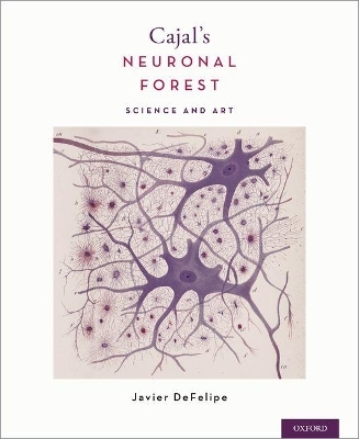
Cajal's Neuronal Forest
Oxford University Press Inc (Verlag)
9780190842833 (ISBN)
This book has been divided into two parts. The first focuses on the scientific atmosphere in Cajal's times, on the history of the neuron, and the anatomical challenge posed in studying neuronal connections. It also delves into the artistic skills of Cajal and other important pioneers in neuroscience and how the neuronal forests have served as an unlimited source of artistic inspiration. The second consists of 275 original drawings by Cajal. All were published over the course of his scientific career and cover virtually all of his research fields of interest, including the spinal cord, the optic lobe and retina, cerebral cortex, and many other regions of the brain.
Cajal's Neuronal Forest: Science and Art is a testament to the natural beauty found in science. Despite the common misconception that the drawings of Cajal and other scientists of the time are pieces of art, these drawings are in fact copies of histological preparations and contributed greatly to the discoveries made in the field of neuroscience. This book is a gem in any library, whether serving as a medical history or a gallery of stunning sketches.
Javier DeFelipe is a Research Professor at the Instituto Cajal (CSIC), Madrid, Spain. His main area of expertise lies in the microanatomy of the cerebral cortex. The frequent citation of his work in the scientific literature reflects the variety and excellence of his numerous contributions. Prof. DeFelipe is also fascinated by the link between the study of the brain and art and has been involved in organizing and curating numerous exhibitions around the world. He has written several articles, chapters and books on both this subject and the influence of Santiago Ramón y Cajal in modern neuroscience.
Table of Contents
Foreword Ramon Areces Foundation
Foreword Spanish Foundation to Science and Technology
Foreword Tatiana Perez De Guzman El Bueno Foundation
Foreword by Pasko Rakic
Preface
Part I: Introduction
Introductory Remarks
A Note on Microphotography and Illustrations of Histological Preparations
The Anatomical Challenge of the Study of Neuronal Connections
Section A: The Scientific Atmosphere in Cajal's Times
The Discovery of the Reazione Nera: Commencing the Detailed Study of the Structure of the Nervous System
The Discovery of the Golgi Method by Cajal and the Influence of Maestre De San Juan and Simarro
Drawing as a Tool to Illustrate Microscopic Images
Differences in the Interpretation of the Microscopic World
Neuron Doctrine versus the Reticular Theory
The Reticular Theory and Precursors of the Neuron Theory
Golgi, Cajal and the Nobel Prize Lectures in 1906
The Law of Dynamic Polarization of Nerve Cells
Introduction of the Term "Neuron" and the Renascence of Reticularism
Confirmation of the Free Ending of the Nervous Processes: The Discovery
of the Synaptic Cleft
Neoreticularism and the Neuron Doctrine in the Present Day
Dendritic Spines: True Anatomical Structures versus Artifact
Dendritic Spines and the Methylene Blue Method
Other Interpretations of the Dendritic Spines
Dendritic Spines and Cajal's 'Final Lesson'
Confirmation that Dendritic Spines are Postsynaptic Structures
Combination of the Golgi Method and Electron Microscopy
Dendritic Spines: Their Features and Role as a Key Component in Microcircuits
Section B: The Golden Era for Artistic Creativity in Neuroscience
Artistic Skills and Emotions of Cajal and Other Early Neuroanatomists
Verification of the Drawings of Cajal and Other Scientists
The Neuronal Forest as a Source of Artistic Inspiration
Summary, Final Considerations and Concluding Remarks
Notes
Bibliography
Part II: Gallery of Cajal's Drawings
Section A: Spinal Cord
Section B: Medulla Oblongata
Section C: Cerebellum and Deep Cerebellar Nuclei
Section D: Midbrain and Thalamus
Section E: Optic Lobe of Lower Vertebrates
Section F: Retina of Vertebrates
Section G: Cerebral Cortex and Olfactory Apparatus
Section H: Degeneration and Regeneration of the Nervous System
Section I: Retina and Optic Centers of Cephalopods
Section J: Insect Brain: Visual System
Section K: Supplementary Figures
Author Index
| Erscheinungsdatum | 29.01.2018 |
|---|---|
| Verlagsort | New York |
| Sprache | englisch |
| Maße | 229 x 282 mm |
| Gewicht | 2059 g |
| Themenwelt | Kunst / Musik / Theater ► Malerei / Plastik |
| Medizin / Pharmazie ► Medizinische Fachgebiete ► Neurologie | |
| Studium ► 1. Studienabschnitt (Vorklinik) ► Anatomie / Neuroanatomie | |
| Studium ► 1. Studienabschnitt (Vorklinik) ► Physiologie | |
| Naturwissenschaften ► Biologie ► Humanbiologie | |
| Naturwissenschaften ► Biologie ► Zoologie | |
| ISBN-13 | 9780190842833 / 9780190842833 |
| Zustand | Neuware |
| Informationen gemäß Produktsicherheitsverordnung (GPSR) | |
| Haben Sie eine Frage zum Produkt? |
aus dem Bereich


