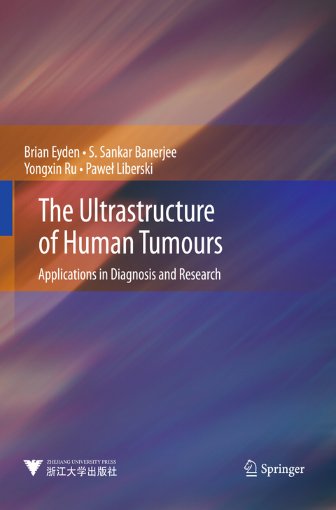
The Ultrastructure of Human Tumours
Springer Berlin (Verlag)
978-3-642-39167-5 (ISBN)
The Ultrastructure of Human Tumours: Applications in Diagnosis and Research describes the core features as seen by transmission electron microscopy, defining the different types of cellular differentiation in tumours; this is relevant for tumour nomenclature and diagnosis, which, in turn, are important for tumour pathologists in their collaboration with oncologists for the treatment of cancer patients. The book is divided into 8 chapters. Following an introduction on technique and procedure, there are chapters on epithelial tumours, melanocytic lesions, soft-tissue and related tumours, lymphoma and leukaemia, CNS neoplasms and neuroendocrine and neuronal tumours. Each chapter includes an introductory text that puts the ultrastructural features in the context of classical pathology. The book includes many new findings and interpretations from well-known tumours, as well as ultrastructural information on several newly described tumour entities not dealt with in existing tumour ultrastructure monographs. The book will especially be of value to tumour pathologists who need to solve problem cases with the aid of electron microscopy, but also to cancer research and tissue engineering scientists working to develop anti-cancer and stem-cell-based therapies. However, even those without access to electron microscopy may also benefit from this book, since many of the images provide an 'explanation' of the appearances of cells, tissues and tumours familiar to pathologists and scientists from light microscopy. In this respect, it is hoped that this book will stimulate the wider use of electron microscopy in pathology. The book is comprehensively referenced, 680 pages long and lavishly illustrated with 757 figures.
Dr. Brian Eyden is Consultant Clinical Scientist and Dr. S. Sankar Banerjee Consultant Histopathologist in the Department of Histopathology, Christie NHS Foundation Trust, Manchester, UK; Dr. Yongxin Ru is Director of theDepartment of Electron Microscopy, Institute of Hematology and Blood Diseases Hospital, Tianjin, China; Pawel Liberski is Professor in the Department of Molecular Pathology and Neuropathology, Medical University Lódz, Poland.
Epithelial Tumours, Especially Carcinoma Squamous and Glandular Differentiation.- Malignant Melanoma.- Soft-Tissue/Mesenchymal/Spindle Cell Tumours.- Tumours of the Nervous System.- Haematolymphoid Neoplasia.- Tumours of Complex or Uncertain Differentiation.
| Erscheint lt. Verlag | 6.3.2014 |
|---|---|
| Zusatzinfo | XVI, 680 p. 45 illus., 28 illus. in color. |
| Verlagsort | Berlin |
| Sprache | englisch |
| Maße | 210 x 279 mm |
| Gewicht | 2065 g |
| Themenwelt | Medizin / Pharmazie ► Medizinische Fachgebiete ► Onkologie |
| Studium ► 2. Studienabschnitt (Klinik) ► Pathologie | |
| Naturwissenschaften ► Physik / Astronomie ► Angewandte Physik | |
| Schlagworte | ATSTC • Biological Microscopy • Cancer Research • electron microscopy • Pathology • Tumor • tumour diagnosis • ZJUP |
| ISBN-10 | 3-642-39167-2 / 3642391672 |
| ISBN-13 | 978-3-642-39167-5 / 9783642391675 |
| Zustand | Neuware |
| Haben Sie eine Frage zum Produkt? |
aus dem Bereich


