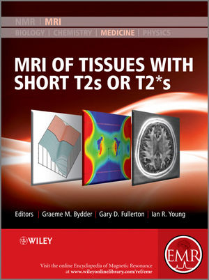
MRI of Tissues with Short T2s or T2*s
John Wiley & Sons Inc (Verlag)
978-0-470-68835-9 (ISBN)
- Titel z.Zt. nicht lieferbar
- Versandkostenfrei
- Auch auf Rechnung
- Artikel merken
Up to now MRI could not be used clinically for imaging fine structures of bones or muscles. Since the late 1990s however, the scene has changed dramatically. In particular, Graeme Bydder and his many collaborators have demonstrated the possibility – and importance – of imaging structures in the body that were previously regarded as being “MR Invisible”. The images obtained with a variety of these newly developed methods exhibit complex contrast, resulting in a new quality of images for a wide range of new applications.
This Handbook is designed to enable the radiology community to begin their assessment of how best to exploit these new capabilities. It is organised in four major sections – the first of which, after an Introduction, deals with the basic science underlying the rest of the contents of the Handbook. The second, larger, section describes the techniques which are used in recovering the short T2 and T2* data from which the images are reconstructed. The third and fourth sections present a range of applications of the methods described earlier. The third section deals with pre-clinical uses and studies, while the final section describes a range of clinical applications. It is this last section that will surely have the biggest impact on the development in the next few years as the huge promise of Short T2 and T2* Imaging will be exploited to the benefit of patients.
In many instances, the authors of an article are the only research group who have published on the topic they describe. This demonstrates that this Handbook presents a range of methods and applications with a huge potential for future developments.
About EMR Handbooks / eMagRes Handbooks
The Encyclopedia of Magnetic Resonance (up to 2012) and eMagRes (from 2013 onward) publish a wide range of online articles on all aspects of magnetic resonance in physics, chemistry, biology and medicine. The existence of this large number of articles, written by experts in various fields, is enabling the publication of a series of EMR Handbooks / eMagRes Handbooks on specific areas of NMR and MRI. The chapters of each of these handbooks will comprise a carefully chosen selection of articles from eMagRes. In consultation with the eMagRes Editorial Board, the EMR Handbooks / eMagRes Handbooks are coherently planned in advance by specially-selected Editors, and new articles are written (together with updates of some already existing articles) to give appropriate complete coverage. The handbooks are intended to be of value and interest to research students, postdoctoral fellows and other researchers learning about the scientific area in question and undertaking relevant experiments, whether in academia or industry.
Have the content of this Handbook and the complete content of eMagRes at your fingertips!
Visit: www.wileyonlinelibrary.com/ref/eMagRes
View other eMagRes publications here
Graeme M Bydder is professor of radiology specializing in magnetic resonance imaging. He has published articles on magnetic resonance techniques, clinical applications of magnetic resonance, image interpretation and related subjects. He is the main developer of UTE. Professor Ian Young, from Marlborough, who helped develop the Magnetic Resonance Imaging (MRI) technology,?has received the Sir Frank Whittle Medal. MRI uses special imaging techniques to take pictures of inside the body. Professor Young, 72, was one of two authors who published the first MRI-generated image of a head in 1978. He also built the world's first MRI machine to use a super-conducting magnet for imaging, an approach now in almost universal use.
Contributors ix
Series Preface xvii
Preface xix
Part A: Basic Science 1
1 An Introduction to Short and Ultrashort T2/T2* Echo Time (UTE) Imaging
Ian Young 3
2 The Physics of Relaxation
John C. Gore, Adam W. Anderson 15
3 Mechanisms for Short T2 and T2* in Collagen-Containing Tissue
Lada V. Krasnosselskaia 31
4 Physical Chemistry of Collagen: The Molecular Basis of Magic Angle Contrast
Gary D. Fullerton 43
Part B: Techniques 59
5 Centric SPRITE MRI of Biomaterials with Short T2*s
Igor V. Mastikhin, Bruce J. Balcom 61
6 Selective Excitation for Ultrashort Echo Time Imaging
John M. Pauly 69
7 Practical Implementation of UTE Imaging
Paul M. Margosian, Tetsuhiko Takahashi, Masahiro Takizawa 79
8 MRI with Zero Echo Time
M. Weiger, K. P. Pruessmann 97
9 AWSOS Pulse Sequence and High-Resolution UTE Imaging
Yongxian Qian, Fernando E. Boada 111
10 Capturing Signals from Fast-Relaxing Spins with Frequency-Swept MRI: SWIFT
Michael Garwood, Djaudat Idiyatullin, Curtis A. Corum, Ryan Chamberlain, Steen Moeller, Naoharu Kobayashi, Lauri J. Lehto, Jinjin Zhang, Robert O’Connell, Michael Tesch, Mikko J. Nissi, Jutta Ellermann, Donald R. Nixdorf 125
11 Imaging in the Presence of Prostheses
Brian A. Hargreaves, Pauline W. Worters, Kim Butts Pauly, John M. Pauly, Garry E. Gold, Kevin M. Koch 143
12 MR Imaging near Metal with UTE–MAVRIC Sequences
Michael Carl, Kevin M. Koch, Jiang Du 155
13 Effects of Hip Prostheses In Situ Exposed to 64 and 128 MHz RF Fields
Jeffrey W. Hand, Donald W. McRobbie 163
14 Absorption Methods for ESR and NMR Imaging of Solid Materials
Andrew J. Fagan, David J. Lurie 171
Part C: Preclinical 185
15 Contrast Manipulation in MR Imaging of Short T2 and T2* Tissues
Nikolaus M. Szeverenyi, Michael Carl 187
16 Magnetization Transfer – Ultrashort Echo Time (MT-UTE) Imaging
Fabian Springer, Petros Martirosian, Fritz Schick 197
17 Ultrashort TE Phase and Spectroscopic Imaging of Short T2 Tissues in the Musculoskeletal System
Jiang Du, Michael Carl, Graeme M. Bydder 209
18 Quantitative Ultrashort TE (UTE) Imaging of Short T2 Tissues
Jiang Du 221
19 MRI-Based Attenuation Correction for Emission Tomography Using Ultrashort Echo Time Sequences
Vincent Keereman, Christian Vanhove, Stefaan Vandenberghe 235
20 Imaging of Very Fast Flows with PC-UTE
Kieran R. O’Brien, Matthew D. Robson 249
21 Double-Quantum Filtered MRI of Connective Tissues
Gil Navon, Uzi Eliav 261
22 Positive-Contrast Visualization of Iron-Oxide-Labeled Cells
Peter M. Jakob, Daniel Haddad 273
Part D: Clinical 287
23 Imaging of Short and Ultrashort T2 and T2* Components of Tissues, Fluids and Materials in the Body Using Clinical Magnetic Resonance Systems
Graeme M. Bydder 289
24 Image-Based Assessment of Cortical Bone
Felix W. Wehrli 305
25 Ultrashort Echo Time Imaging of Phosphorus in Man
Matthew D. Robson 319
26 Knee
Emily J. McWalter, Hillary J. Braun, Kathryn E. Keenan, Garry E. Gold 325
27 Short and Ultrashort TE Imaging of Cartilage and Fibrocartilage
Won C. Bae, Eric Y. Chang, Christine B. Chung 339
28 Myelin Water Imaging
Alex L. MacKay, Cornelia Laule 359
29 Quantitative Metabolic MR Imaging of Human Brain Using 17O and 23Na
Ian C. Atkinson, Aiming Lu, Keith R. Thulborn 377
30 Sodium MRI in Man: Technique and Findings
Paul A. Bottomley 397
31 Short T2/T2* Imaging of Calcification and Atherosclerosis
Sonia Nielles-Vallespin 415
32 Ultrashort TE in Cancer Imaging
Konstantina Boulougouri, Christina Messiou, Nandita M. deSouza 425
33 Ultrashort TE Imaging of Cryotherapy
Aiming Lu, Bruce L. Daniel, Kim Butts Pauly 433
34 Imaging around Orthopedic Hardware: Clinical Applications
Catherine L. Hayter, Hollis G. Potter 449
Index 463
| Erscheint lt. Verlag | 4.1.2013 |
|---|---|
| Reihe/Serie | eMagRes Books |
| Verlagsort | New York |
| Sprache | englisch |
| Maße | 193 x 254 mm |
| Gewicht | 1388 g |
| Themenwelt | Medizin / Pharmazie ► Medizinische Fachgebiete ► Radiologie / Bildgebende Verfahren |
| Naturwissenschaften ► Chemie | |
| ISBN-10 | 0-470-68835-1 / 0470688351 |
| ISBN-13 | 978-0-470-68835-9 / 9780470688359 |
| Zustand | Neuware |
| Haben Sie eine Frage zum Produkt? |
aus dem Bereich


