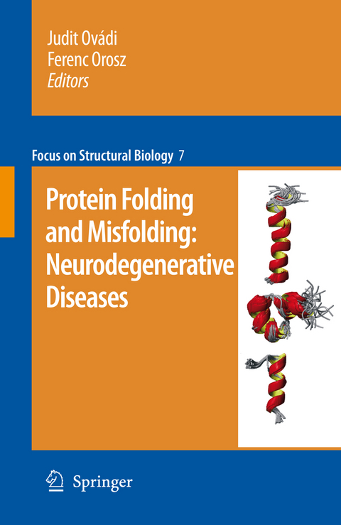
Protein folding and misfolding: neurodegenerative diseases
Springer (Verlag)
978-90-481-8127-8 (ISBN)
It was twenty ?ve years ago this year that for the ?rst time a protein under- ing a form of human cerebral amyloidosis, the Icelandic-type hereditary cerebral haemorrhage was identi?ed. This, together with the recognition that an amino acid substitution can transform the wild type cystatin C into a disease-associated amyloid-forming protein in this condition, was only a prelude to a series of imp- tant discoveries that followed. As a result, pathologically altered proteins have been brought into the centre stage of research into the pathomechanism of a n- ber of neurodegenerative diseases, which include epidemiologically such important conditions as Alzheimer's disease or Parkinson's disease and, among others, also the transmissible spongiform encephalopathies, Huntington's chorea, spinocereb- lar ataxias, frontotemporal lobar degenerations and amyotrophic lateral sclerosis.
Despite the diversity in the amino acid sequence of the different proteins involved in these neurological diseases, one of the common themes underlying the patho- chanisms of all these conditions is protein misfolding, aggregation - hence the term protein folding disorders -, which can trigger cascades of events ultimately resulting in synapse loss and neuron death with devastating clinical consequences in many of the most precious spheres of human existence including personality, cognition, memory, skilled movements and affection. It is always a challenging task to unite the different topics of the individual ch- ters into a common theme in a multi-author volume, but the current book edited by Judit Ovadi and Ferenc Orosz ?ts this task admirably.
1. Structural Disorder and Its Connection with Misfolding Diseases; Veronika Csizmok and Peter Tompa. 1.1 The Concept of Protein Disorder. 1.2 Biophysical and Bioinformatics Characterization of Disorder. 1.2.1 Biophysical Techniques 1.2.2 Bioinformatics Techniques. 1.3 Disorder in Vivo, the Effect of Crowding? 1.4 Disorder and Aggregation. 1.5 Disorder in Neurodegenerative Diseases. 1.6 Physiological Prions. 1.7 Structural Transition to Amyloid: Partially Folded Intermediates. 1.8 The Structure of Amyloid: Cross-Beta Models and Flexibility. 1.9 Conclusions. References. 2 Intrinsic Disorder in Proteins Associated with Neurodegenerative Diseases Vladimir N. Uversky. 2.1 Neurodegenerative Diseases as Proteinopathies. 2.2 Introducing Intrinsically Disordered Proteins. 2.2.1 Concept. 2.2.2 Experimental Techniques for IDP Detection. 2.2.3 Sequence Peculiarities of IDPs and Predictors of Intrinsic Disorder. 2.2.4 Abundance of IDPs and their Functions. 2.3Abundance of IDPs in Neurodegenerative Diseases. Evidence from the Bioinformatics Analyses. 2.4 Intrinsic Disorder in Proteins Associated with -Protein and Alzheimer’s Disease. 2.4.2 Neurodegenerative Diseases. 2.4.1 Amyloid Tau Protein in Alzheimer’s Disease and Other Tauopathies. 2.4.3 Prion Protein and Prion Diseases. 2.4.4 Synucleins - and Synuclein and Synucleinopathies. 2.4.5 Parkinson’s Disease and Dementia with Lewy Bodies. 2.4.6 Polyglutamine Repeat Diseases and Huntingtin, Ataxin-1, Ataxin-3, androgen Receptor and Atrophin-1. 2.4.7 Abri Peptide and Familial British Dementia. 2.4.8 Adan in Familial Danish Dementia. 2.4.9 Glial Fibrillary Acidic Protein and Alexander and Alpers Disease. 2.4.11DNA Disease. 2.4.10 Mitochondrial DNA Polymerase Excision Repair Protein ERCC-6 and Cockayne Syndrome. 2.4.12 Survival of Motor Neurons Protein and Spinal Muscular Atrophy. 2.5 Concluding Remarks: Another Illustration of the D2 Concept. References. 3 Dynamic Role of Ubiquitination in the Management of MisfoldedProteins Associated with Neurodegenerative Diseases. Esther S.P. Wong, Jeanne M.M. Tan and Kah-Leong Lim. 3.1 Protein Misfolding and the Ubiquitin-Proteasome System. 3.2 Protein Misfolding, UPS Disruption and Neurodegeneration. 3.3 Diversity of Ubiquitin Modifications. 3.4 Non-Proteolytic Ubiquitination and Protein Inclusions Biogenesis. 3.5 Aggresomes Formation and Clearance. 3.6 K63-Linked Polyubiquitination – A Novel Cargo Recognition Signal For Autophagic Degradation. 3.7 A Model of Inclusion Biogenesis and Clearance. 3.8 E2/E3 Pairs – Triage officers?. 3.9 Conclusions. References. 4. Protein Misfolding and Axonal Protection in Neurodegenerative Diseases. Haruhisa Inoue, Takayuki Kondo and Ryosuke Takahashi. 4.1 Neuronal Dysfunction in Neurodegeneration Are Reversible Process. 4.2 Neuronal Dysfunction Is Not Treatable by Anti-Cell Death Therapy. 4.3 Morphological Aspects of Neuronal Dysfunction Caused by Protein Aggregation/Misfolding in Human Neurodegenerative Disorder. 4.4 Protein Misfolding and Axonal Degeneration in Experimental Animal Models. 4.5 Therapeutic approaches to treat neuronal dysfunction by axonal protection. 4.5.1 Axonal regeneration. 4.5.2 Anti-Wallerian Degeneration. 4.5.3 Autophagy Enhancement. 4.5.4 Stabilization of Microtubules. 4.6 Concluding remarks. References. 5. Endoplasmic Reticulum Stress in Neurodegeneration Jeroen J.M. Hoozemans and Wiep Scheper. 5.1Introduction. 5.2 Protein Quality Control in the Endoplasmic Reticulum. 5.2.2 Triage: ERAD. 5.2.3 Degradation: Ubiquitin Proteasome System and Autophagy. 5.2.4 Stress Response: The Unfolded Protein Response. 5.2.5 ER-Stress-Induced Cell Death. 5.3. ER Stress in Neurodegenerative Disorders. 5.3.1 Alzheimer’s Disease. 5.3.2 Parkinson’s Disease 5.3.3 Prion Disease. 5.3.4 Tauopathies. 5.3.5 Polyglutamine Diseases. 5.3.6 Amyotrophic Lateral Sclerosis. 5.3.7 White Matter Disorders. 5.4 Conclusions .References. 6. Involvement of Alpha-2 Domain in Prion Protein Conformationally-InducedDiseases Luisa Ronga, Pasquale Palladino, Ettore Benedetti, Raffaele Ragone and Filomena Rossi. 5.1. Conformational Diseases. 5.2. Prion Biology. 5.3. Approaches to TSE therapy: Anti-Prion Compounds. 5.4. Immune Intervention. 5.5. Prion Protein Structure. 5.6. Determinants of Prpc Conversion: the N-Terminal Region. 5.7. Determinants of Prpc Conversion: the C-Globular Domain. 5.8. Prion-Metal Ion Binding. 5.9. The a -2 Helix Domain: What Role?. 5.10. Solution Structure of Prp[173-195] and Its Analogues. 5.11. Metal Ion Titration. 5.12. Anion-Induced Effects. 5.13. Conclusions. References. 7 Synuclein Structure and Function in Parkinson's Disease. David Eliezer. 7.1 Background. 7.1.1 The Discovery of Synucleins. 7.1.2 Synuclein Mutations in Parkinson’s Disease. 7.1.3 Synuclein in Lewy Bodies. 7.1.4 Synuclein Toxicity. 7.1.5 Synuclein Function. 7.2 Free State Structure. 7.2.1 Residual Secondary Structure. 7.2.2 Role of Residual Structure in Aggregation. 7.2.3 Transient Long-Range Interactions. 7.3 Fibril Structure. 7.4 Lipid-Bound Structure. 7.4.1 A Role for Synuclein Function in Disease?. 7.4.2 Secondary Structure. 7.4.3 Topology. 7.4.4 Effects of Parkinson’s Disease-Linked Mutations. 7.5Synuclein. 7.6 New Model for Synuclein Function. References. 8 Inhibition of a-Synuclein Aggregation by Antioxidants and Chaperones in Parkinson’s Disease Jean-Christophe Rochet and Fang Liu. 8.1 Introduction. 8.2 Molecular Details of a-Synuclein Aggregation. 8.2.1 a-Synuclein Is a Natively Unfolded, Presynaptic Protein. 8.2.2 a-Synuclein Forms Fibrils and Protofibrils. 8.2.3 a-Synuclein Protofibrils Permeabilize Membranes. 8.2.4 a-Synuclein Self-Assembly Is Promoted by Membranes. 8.2.5 a-Synuclein Aggregates Disrupt Protein Clearance Mechanisms. 8.2.6 a-Synuclein Is Post-Translationally Modified in Parkinson’s Disease. 8.2.7 a-Synuclein Self-Assembly Is Stimulated by Oxidative Modifications. 8.2.8 a-Synuclein Aggregation Is Modulated by Phosphorylation. 8.2.9 Summary (PartI). 8.3 Inhibition of a-Synuclein Aggregation by Molecules with Antioxidant Activity. 8.3.1 Inhibition of a-Synuclein Aggregation by MsrA. 8.3.2 Inhibition of a-Synuclein Aggregation by Small-Molecule Antioxidants. 8.3.3 Summary (Part II). 8.4 Inhibition of a-Synuclein Aggregation by Molecular Chaperones. 8.4.1 Inhibition of a-Synuclein Aggregation by Hsp70. 8.4.2 Inhibition of a-Synuclein Aggregation by aB-Crystallin and Hsp27. 8.4.3 Interaction Between a-Synuclein and Hsp90. 8.4.4 Inhibition of a-Synuclein Aggregation by TorsinA. 8.4.5 Inhibition of a-Synuclein Aggregation by DJ-1. 8.4.6 -Synucleinopathies Summary (Part III). 8.5 Concluding remarks. References. 9 Novel Proteins in Christine Lund Petersen and Poul Henning Jensen. 9.1 Introduction. 9.2 The Synucleinopathies. 9.3 -Synuclein Aggregation. 9.4.1 Synuclein. 9.4 Proteins Involved in Brain-Specific Protein P25a/TPPP. 9.4.2 Tau. 9.4.3 Synphilin-1. 9.4.4 TAR-DNA-Binding Protein 43 (TDP-43). 9.4.5 Leucine-Rich Repeat Kinase 2 (LRRK2). 9.4.6 FK506-Binding Proteins. 9.4.7 Histones 9.4.8 Other -Synuclein Aggregation. 9.5 Concluding Remarks. References. Proteins Involved in 10 TPPP/P25: A New Unstructured Protein Hallmarking Synucleinopathies Ferenc Orosz, Attila Lehotzky, Judit Olah Stb and Judit Ovádi. 10.1 Occurrence of TPPP Proteins. 10.2 Unfolded Structural Features of TPPP Proteins. 10.2.1 Prediction. 10.2.2 Experimental. 10.3 Interacting Partners of TPPP/P25. 10.4 TPPP Expression At Cell Level. 10.4.1 Structures and Effects in Living Cells. 10.4.2 Energy State. 10.4.3 Function. 10.4.4 Human Cell Model For Aggresome Development. 10.5 TPPP/P25 in Brain Tissues. 10.5.1 Normal Brain. 10.5.2 Pathological Brain. 10.6 Impact of Unfolded Structures On Physiological and Pathological Functions. References. 11 Tau protein in Alzheimer's Disease and Other Tauopathic Neurodegenerative Diseases Jeff Kuret. 12 Protein-Base Neuropathology and Molecular Classification of Human Neurodegenerative Diseases Gabor G. Kovacsand Herbert Budka. 12.1 Introduction. 12.2 Classification of Neurodegenerative Disease: Basic Concepts. 12.3 Proteins with Relevance For the Classification of Neurodegenerative Diseases. 12.3.1 Microtubule-Associated Protein Tau. 12.3.2 b -Amyloid. 12.3.3 a -Synuclein. 12.3.4 Prion Protein. 12.3.5 TAR-DNA-Binding Protein 43 (TDP-43). 12.3.6 a -Internexin. 12.3.7 Other Proteins. 12.4 Morphological Types of Extra- and Intracellular Protein Deposits. 12.4.1 Extracellular Protein Deposition. 12.4.2 Intracellular Protein Deposition. 12.5 Other Distinguishing Morphological Features. 12.6 Synthesis: Classification of Neurodegenerative Diseases. 12.7 Concluding Remarks. References. Index
| Erscheint lt. Verlag | 28.10.2010 |
|---|---|
| Reihe/Serie | Focus on Structural Biology ; 7 |
| Zusatzinfo | XIV, 278 p. |
| Verlagsort | Dordrecht |
| Sprache | englisch |
| Maße | 155 x 235 mm |
| Themenwelt | Studium ► 1. Studienabschnitt (Vorklinik) ► Physiologie |
| Naturwissenschaften ► Biologie ► Biochemie | |
| Naturwissenschaften ► Biologie ► Humanbiologie | |
| Naturwissenschaften ► Biologie ► Zellbiologie | |
| Naturwissenschaften ► Biologie ► Zoologie | |
| ISBN-10 | 90-481-8127-5 / 9048181275 |
| ISBN-13 | 978-90-481-8127-8 / 9789048181278 |
| Zustand | Neuware |
| Haben Sie eine Frage zum Produkt? |
aus dem Bereich


