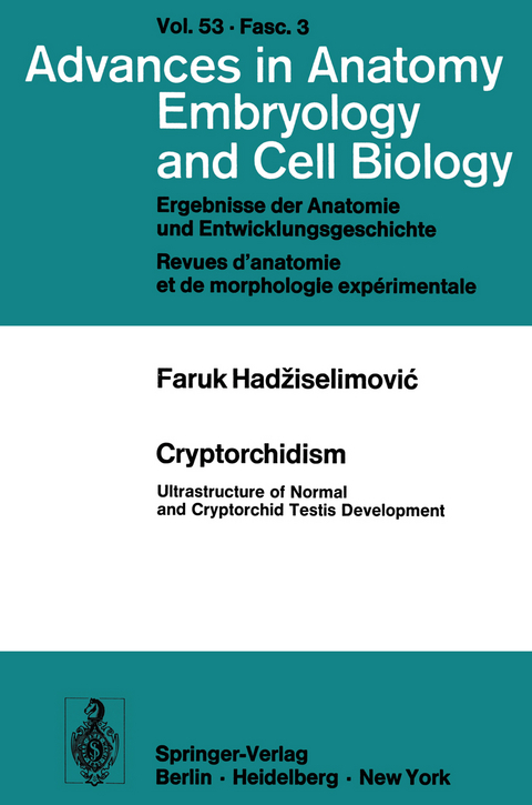
Cryptorchidism
Ultrastructure of Normal and Cryptorchid Testis Development
Seiten
1977
Springer Berlin (Verlag)
978-3-540-08361-0 (ISBN)
Springer Berlin (Verlag)
978-3-540-08361-0 (ISBN)
Cytological techniques have greatly improved in the last twenty years, largely as a result of further development of the microscope. Electron microscopy, in particular, has opened up great prospects for the study of cell morphology, while the develop ment of radio-immuno-assay has brought great progress in endocrinology. The applica tion of these two techniques, which are complementary to each other and provide mutually supporting evidence, is the subject of this monograph. The work is divided into two parts. The first deals with the ultrastructure of normal testicular development, describing four main elements of the testicle - the germ cells, the Sertoli cells, the peri tubular connective tissue and the Leydig cell- with details of their individual development. To make the electron micrographs more easily understandable, diagrams have been used to explain the most important points. The second part deals with cryptorchid testicles, including the primary and sec ondary changes involved and morphometric studies of the secondary changes. The significance of these ultrastructural observations for the treatment of cryptorchidism is emphasized. With the aid of radio-immuno-assay the level of testosterone in cryp torchid mice was determined and comparisons drawn between the ultrastructural changes in cryptorchidism in mouse and man. The experimental studies served as a basis for the hypothesis that, in all probability, a congenital disturbance of the hy pothalamo-hypophyso-gonadal axis is responsible for cryptorchidism. I hope that this monograph will contribute towards a better understanding of normal and cryptorchid testicular development and of the etiology of cryptorchidism.
Preface.- Material and Methods.- 1. Biopsies from Child Testicles.- 2. Animal Experiments.- The Normal Testicle.- 1. General Observations on the Development of the Normal Testicle in Children.- 2. Germ Cells.- 3. Sertoli Cells.- 4. Peritubular Connective Tissue.- 5. LeydigCells.- Cryptorchidism.- 1. Germ Cells.- 2. Sertoli Cells.- 3. Peritubular Connective Tissue.- 4. Leydig Cells.- 5. Estrogen Induced Cryptorchidism in Mice.- Discussion.- Therapy.- Programme of Therapy.- Summary.- Acknowledgements.- References.
| Erscheint lt. Verlag | 1.8.1977 |
|---|---|
| Reihe/Serie | Advances in Anatomy, Embryology and Cell Biology |
| Zusatzinfo | 72 p. 23 illus. |
| Verlagsort | Berlin |
| Sprache | englisch |
| Maße | 155 x 235 mm |
| Gewicht | 160 g |
| Themenwelt | Medizin / Pharmazie ► Medizinische Fachgebiete ► Urologie |
| Naturwissenschaften ► Biologie | |
| Schlagworte | biopsy • Cell • Development • electron microscopy • endocrinology • Microscopy • Morphology • Testis • tissue |
| ISBN-10 | 3-540-08361-8 / 3540083618 |
| ISBN-13 | 978-3-540-08361-0 / 9783540083610 |
| Zustand | Neuware |
| Haben Sie eine Frage zum Produkt? |
Mehr entdecken
aus dem Bereich
aus dem Bereich


