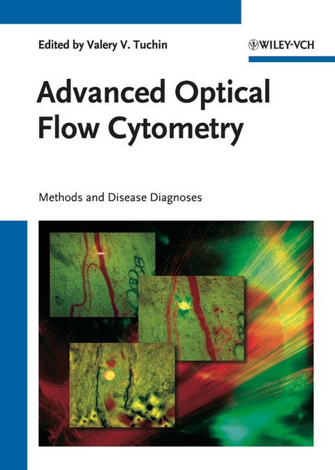
Advanced Optical Flow Cytometry
Wiley-VCH (Verlag)
9783527409341 (ISBN)
- Titel ist leider vergriffen;
keine Neuauflage - Artikel merken
.
A detailed look at the latest research in non-invasive in vivo cytometry and its applications, with particular emphasis on novel biophotonic methods, disease diagnosis, and monitoring of disease treatment at single cell level in stationary and flow conditions. This book thus covers the spectrum ranging from fundamental interactions between light, cells, vascular tissue, and cell labeling particles, to strategies and opportunities for preclinical and clinical research. General topics include light scattering by cells, fast video microscopy, polarization, laser-scanning, fluorescence, Raman, multi-photon, photothermal, and photoacoustic methods for cellular diagnostics and monitoring of disease treatment in living organisms. Also presented are discussions of advanced methods and techniques of classical flow cytometry.
Valery Tuchin is Head of Chair of Optics and Biomedical Physics and Director of Research-Educational Institute of Optics and Biophotonics at Saratov State University. He has authored more than 250 papers and books, including his latest, Tissue Optics. Light Scattering Methods and Instrumentation for Medical Diagnosis (SPIE Tutorial Texts in Optical Engineering, Vol. TT38, 2000; second edition, PM166, 2007), Handbook of Optical Biomedical Diagnostics (SPIE Press, Vol. PM107, 2002), Coherent-Domain Optical Methods for Biomedical Diagnostics, Environmental and Material Science, Kluwer Academic Publishers, Boston, USA, vols. 1 & 2, 2004, Optical Clearing of Tissues and Blood (SPIE Press, Vol. PM154, 2005), and Optical Polarization in Biomedical Applications (co-authors L. Wang and D.A. Zimnyakov; Springer, 2006). Some of the contributors: Martin Leahy, University of Limerick, Ireland Attila Tárnok, University of Leipzig, Germany Andreas O.H. Gerstner, University of Bonn, Germany Anja Mittag, University of Leipzig, Germany Megha Makam, Daisy Diaz, Rabindra Tirouvanziam, Stanford University School of Medicine, USA Steven Boutrus, Derrick Hwu & Cherry Greiner, Tufts University, MA, USA Michael Chan & Charlotte Kuperwasser, Tufts-New England Medical Center, MA, USA Charles P. Lin & Irene Georgakoudi, Harvard Medical School,MA, USA E.I. Galanzha, Saratov State University, Russia V.P. Zharov, Arkansas University of Medical Science, USA A.V. Priezzhev, A.G. Lugovtsov, S.Yu. Nikitin & Yu.I. Gurfinkel, Moscow State University, Russia Valeri P. Maltsev, Maxim A. Yurkin & Elena Eremina,Institute of Chemical Kinetics and Combustion, Novosibirsk, Russia Alfons G. Hoekstra & Thomas Wriedt,University of Amsterdam, The Neverlands Péter Nagy % György Vereb, János Szöllsi, University of Debrecen, Hungary
- Perspectives in Cytometry
- Novel Concepts and Requirements in Cytometry
- Optical Imaging of Cells with Gold Nanoparticle Clusters as Light Scattering Contrast Agents: A Finite-Difference Time-Domain Approach to the Modeling of Flow Cytometry Configurations
- Optics of White Blood Cells: Optical Models, Simulations, Experiments
- Optical Properties of Flowing Flood Cells
- Laser Diffraction by the Erythrocytes and Deformability Measurements
- Characterization of Red Blood Cells Rheological and Physiological State using Optical Flicker Spectroscopy
- Digital Holographic Microscopy for Quantitative Live Cell Imaging and Cytometry
- Comparison of Immunophenotyping and Rare Cell Detection by Slide-based Imaging Cytometry and by Flow Cytometry
- Microfluidic Flow Cytometry: Advancements towards Compact, Integrated Systems
- Label-free Cell Classification with Diffraction imaging flow cytometer
- An Integrative Approach for Immune Monitoring of Human Health and Disease by Advanced Flow Cytometry Methods
- Optical Tweezers and Cytometry
- In-vivo image flow cytometry
- Instrumentation for In-Vivo Flow Cytometry; A Sickle Cell Anaemia Case Study
- Advances in Fluorescence-Based In-vivo Flow Cytometry for Cancer Applications
- In-vivo Photothermal and Photoacoustic Flow Cytometry
- Optical Instrumentation for the Measurement of Blood Perfusion, Concentration, Oxygenation and Shape In-vivo
- Blood Flow Cytometry and Cell Aggregation Study with Laser Speckle
- Modifications of Blood Optical Properties during Photodynamic Reactions in-Vitro and In-vivo
| Erscheint lt. Verlag | 12.4.2011 |
|---|---|
| Sprache | englisch |
| Maße | 170 x 240 mm |
| Gewicht | 1533 g |
| Themenwelt | Naturwissenschaften ► Physik / Astronomie ► Angewandte Physik |
| Schlagworte | Angewandte Mikrobiologie • applied microbiology • Bildgebende Systeme u. Verfahren • Bildgebende Verfahren i. d. Biomedizin • biomedical engineering • Biomedical Imaging • Biomedizinische Technik • Biomedizintechnik • Biophysik • Biowissenschaften • Cytometrie • Electrical & Electronics Engineering • Electrical & Electronics Engineering • Elektrotechnik u. Elektronik • Imaging Systems & Technology • Imaging Systems & Technology • Immunologie • immunology • Laser • Life Sciences • Medical & Health Physics • Medical Cell Biology • Medical & Health Physics • Medical Science • Medizin • Medizinische Zellbiologie • Nanobiotechnologie • nanobiotechnology • Nanotechnologie • nanotechnology • Optics & Photonics • Optics & Photonics • Optik u. Photonik • Physics • Physik • Physik in Medizin u. Gesundheitswesen • Zellbiologie |
| ISBN-13 | 9783527409341 / 9783527409341 |
| Zustand | Neuware |
| Informationen gemäß Produktsicherheitsverordnung (GPSR) | |
| Haben Sie eine Frage zum Produkt? |
aus dem Bereich


