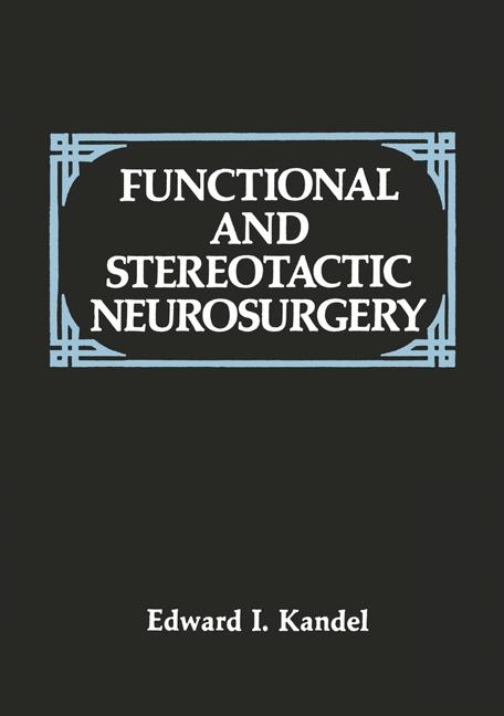
Functional and Stereotactic Neurosurgery
Kluwer Academic/Plenum Publishers (Verlag)
978-0-306-42701-5 (ISBN)
- Titel ist leider vergriffen;
keine Neuauflage - Artikel merken
Foreword.- Preface.- Abbreviations.- 1 Anatomy of Subcortical Structures Related to Stereotaxy.- 1. Extrapyramidal System.- 1.1. Thalamus.- 1.1.1. Ventrolateral Nucleus.- 1.1.2. Posterior Ventrolateral and Ventromedial Nuclei.- 1.1.3. Centrum Medianum.- 1.1.4. Pulvinar.- 1.2. Capsula Interna.- 1.3. Subthalamic Region.- 1.4. Globus Pallidus.- 1.5. Striatum.- 1.5.1. Nucleus Caudatus 4.- 1.5.2. Putamen.- 1.6. Substantia Nigra.- 1.7. Subthalamic Nucleus.- 1.8. Nucleus Ruber.- 2. Limbic System.- 2.1. Amygdaloid Nucleus.- 2.2. Hippocampus.- 2.3. Hypothalamic Region.- 3. Cerebellum.- 4. Pain Structures and Pain-Conducting Pathways of the CNS.- 2 Extrapyramidal Mechanisms.- 1. General Remarks.- 1.1. Regulation of Motor Activity.- 1.2. The Pathogenesis of Extrapyramidal Syndromes.- 2. Tremor.- 2.1. Historical Note.- 2.2. The Characteristics of Tremor.- 2.3. Graphic Recording of Tremor.- 2.4. Physiological Tremor.- 2.5. Pathological Tremor.- 2.6. Action Tremor.- 2.7. Tremor and Proprioceptive Input.- 2.8. Interrelationship of Various Types of Tremor.- 2.9. Pathogenesis of Tremor.- 2.10. Generator of Rhythmic Tremor.- 2.11. Computerized Spectral Analysis of Tremor.- 2.12. Tremor and Cerebral Structures.- 3. Rigidity.- 3.1. Muscle Tone and Its Regulation.- 3.2. Pathogenesis of Extrapyramidal Rigidity.- 4. Spasticity.- 5. Akinesia.- 3 Stereotactic Method.- 1. Historical Note.- 2. General Principles.- 2.1. Cranial Reference Points.- 2.2. Intracerebral Reference Points.- 2.3. Linear Reference Points.- 3. Individual Variability.- 4. Stereotactic Atlases.- 5. Stereotactic Instruments.- 5.1. Stereotactic Frame of Spiegel and Wycis.- 5.2. Stereotactic Frame of Riechert and Mundinger.- 5.3. Stereotactic Frame of Guiot and Gillingham.- 5.4. Stereotactic Frame of Leksell.- 5.5. Stereotactic Frame of Talairach.- 5.6. Stereotactic Frame of Rand and Wells.- 5.7. Stereotactic Frame of Laitinen.- 5.8. Stereotactic Frame of Oliver-Bertrand-Tipal.- 5.9. Stereotactic Frame of Kandel.- 5.10. Other Stereotactic Instruments.- 6. Computer Techniques.- 7. Stereotaxis and Computerized Tomography.- 7.1. Brown-Roberts-Wells Stereotactic System.- 7.2. Patil Stereotactic System.- 8. Stereotaxis and Nuclear Magnetic Resonance.- 9. Stereotaxis and Digital Subtraction Angiography.- 10. Combined Visualization Techniques.- 11. Stereotaxis and Ultrasound.- 4 Principles of Stereotactic Operations on Subcortical Structures 1.- 1. General Considerations.- 2. Anesthesia.- 3. Location of the Burr Hole.- 4. Visualization of the Ventricular System.- 5. Stereotactic Coordinates.- 5.1. Anteroposterior Projection.- 5.2. Lateral Projection.- 6. X-RayControl.- 7. Functional Control.- 7.1. Electrostimulation.- 7.1.1. Ventrolateral Nucleus.- 7.1.2. Sensory Nuclei.- 7.1.3. Pulvinar.- 7.1.4. Centrum Medianum and Intralaminar Nuclei.- 7.1.5. Dorsomedial Nuclei.- 7.1.6. Anterior Nuclei.- 7.1.7. Capsular Interna.- 7.1.8. Subthalamic Region.- 7.1.9. Pallidum (Globus Pallidus).- 7.1.10. Nucleus Caudatus.- 7.1.11. Nucleus Ruber.- 7.1.12. Hypothalamic Region.- 7.1.13. Midbrain Tegmentum.- 7.1.14. Periaqueductal Gray Matter.- 7.1.15. Amygdala.- 7.1.16. Hippocampus.- 7.1.17. Cerebellar Nuclei.- 7.2. Electrosubcorticography.- 7.3. Microelectrode Recording.- 7.4. Evoked Potentials.- 7.5. Impedance Techniques.- 7.6. Other Techniques.- 7.7. Summary of Functional Controls.- 5 The Production of Lesions in Stereotactic Operations.- 1. General Principles.- 2. Local Freezing.- 2.1. Biological Effects of Freezing.- 2.2. Effect of Low Temperatures on Vessels.- 2.3. Experimental Cryoneurosurgery.- 2.4. Factors Determining the Size of the Cryogenic Lesion.- 2.5. Morphological Pathology of Cryogenic Brain Lesions.- 2.6. Cryosurgical Equipment.- 2.7. The Author’s Cryosurgical Instrument.- 2.8. Technique for Producing Cryosurgical Lesions.- 3. Electrical Methods.- 3.1. Anodal Electrolysis.- 3.2. High-Frequency Coagulation.- 3.3. Induction Heating Method.- 4. Radioactive Isotopes.- 5. Laser Techniques.- 6. Remote Control Methods.- 6.1. 7 Irradiation.- 6.2. Proton Beam.- 6.3. Ultrasound.- 7. Chemical Methods.- 8. Mechanical Methods.- 6 Parkinsonism.- 1. Historical Note.- 2. Epidemiology.- 3. Mortality.- 4. Etiology.- 5. Pathological Anatomy.- 6. Pathogenesis.- 7. Clinical Picture.- 7.1 The Beginning and Development of the Disease.- 7.2. Symptomatology.- 7.2.1. Disorders of Posture and Gait.- 7.2.2. Speech Disorders.- 7.2.3. Writing Disorders.- 7.2.4. Vegetative Disorders.- 7.2.5. Pain Syndrome.- 7.2.6. Deformities of the Hands and Feet.- 7.2.7. Mental Disorders.- 7.2.8. Vestibular Disorders.- 7.2.9. Glucose Metabolic Disorders.- 7.2.10. Respiratory Disorders.- 7.2.11. Oculogyric Crises.- 7.2.12. Sleep Disorders.- 7.3. Electroencephalography.- 7.4. Electromyography.- 7.5. Cerebral Blood How.- 7.6. Forms of Parkinsonism.- 7.6.1. Mixed Type.- 7.6.2. Tremor Type.- 7.6.3. Rigid Type.- 7.6.4. Akinetic Type.- 7.7. Stages of Parkinsonism.- 7.8. Diagnosis.- 7.9. Medical Therapy.- 7.9.1. Anticholinergic Agents.- 7.9.2. L-Dopa.- 7.9.3. Dopamine Agonists.- 7.9.4. Amantadine and Deprenyl.- 7.9.5. Postoperative Medication.- 8. Indications and Contraindications for Surgery.- 8.1. Age.- 8.2. Etiology.- 8.3. Form of the Disease.- 8.4. Duration of Disease.- 8.5. State of Internal Organs.- 8.6. Brainstem Functions.- 8.7. Mental State.- 8.8. Hydrocephalus.- 8.9. Medication.- 8.10. Stage of the Disease.- 8.11. Summary.- 8.12. Side of the Operation.- 9. Surgery.- 9.1. Historical Note.- 9.1.1. Operations on the Cerebral Cortex.- 9.1.2. Operations on Subcortical Pathways.- 9.1.3. Frontal Leukotomy.- 9.1.4. Pedunculotomy.- 9.1.5. Bulbotomy.- 9.1.6. Clipping of the Anterior Choroidal Artery.- 9.1.7. Operations on the Cerebellum.- 9.1.8. Operations on the Spinal Cord.- 9.1.9. Operations on the Sympathetic System.- 9.1.10. Operations on the Basal Ganglia.- 9.2. Summary.- 10. Stereotactic Surgery.- 10.1. Stereotactic Destruction of the Subthalamus.- 10.2. Bilateral Operations.- 10.3. Clinicoanatomic Correlations.- 11. Prevention and Treatment of Postoperative Complications.- 12. Personal Observations.- 13. Concluding Remarks.- 7 Dystonia Musculorum Deformans.- 1. Historical Note.- 2. Etiology.- 3. Pathology.- 4. Pathogenesis.- 5. Clinical Investigation.- 5.1. Westphal Phenomenon.- 5.2. Effect of Thalamotomy.- 6. Biochemical Pathogenesis.- 7. Clinical Aspects.- 7.1. Stages of the Disease.- 7.2. Diagnostic Studies.- 7.3. Differential Diagnosis.- 8. Indications and Contraindications for Surgical Treatment.- 9. Surgical Treatment.- 10. Stereotactic Operations.- 11. Personal Observations.- 8 Spasmodic Torticollis.- 1. Introduction.- 2. Etiology.- 3. Pathology.- 4. Pathogenesis.- 5. Clinical Picture.- 6. Stages of the Disorder.- 7. Other Manifestations of ST.- 8. Differential Diagnosis.- 9. Surgical Treatment.- 9.1. Cervical Rhizotomy.- 9.2. Stereotactic Operations.- 9.3. Stimulation of Spinal Cord.- 10. Personal Observations.- 11. Summary.- 9 Cerebral Palsy.- 1. Introduction.- 2. Etiology.- 3. Pathology and Pathogenesis.- 4. Clinical Aspects.- 5. Surgical Treatment.- 5.1. Stereotactic Operations.- 5.1.1. Stereotactic Thalamotomy.- 5.1.2. Stereotactic Destruction of Pulvinar (Pulvinotomy).- 5.1.3. Stereotactic Dentatotomy.- 5.1.4. Stereotactic Pallidotomy.- 5.1.5. Stereotactic Putamenotomy.- 5.1.6. Combined Operations.- 5.2. Selective Posterior Rhizotomy.- 5.3. Stimulation Methods.- 6. Personal Observations.- 10 Hemihyperkinesias.- 1. Introduction.- 2. Etiology and Pathogenesis.- 3. Clinical Features.- 4. Stereotactic Surgery.- 5. Personal Observations.- 11 Multiple Sclerosis.- 1. Introduction.- 2. Surgical Treatment.- 2.1. Stereotactic Operations.- 2.2. Operations on the Spinal Cord.- 3. Personal Observations.- 12 Hereditary Degenerative Disorders.- 1. Introduction.- 2. Hepatocerebral Dystrophy (Wilson’s Disease).- 2.1. Background.- 2.2. Surgical Treatment.- 3. Huntington’s Chorea.- 3.1. Background.- 3.2. Surgical Treatment.- 4. Essential Tremor.- 4.1. Background.- 4.2. Surgical Treatment.- 5. Hunt’s Cerebellar Dyssynergia.- 5.1. Background.- 5.2. Surgical Treatment.- 6. Myoclonus and Myoclonus Epilepsy.- 6.1. Background.- 6.2. Myoclonus.- 6.3. Myoclonus Epilepsy.- 6.4. Surgical Treatment.- 13 Pain.- 1. General Remarks.- 2. Gate Control Theory.- 3. Antinociceptive System.- 4. Pathogenesis of Chronic Pain.- 5. Surgical Management of Pain.- 5.1. General Remarks.- 5.2. Operations on the Brain.- 5.2.1. Operations on Thalamic Nuclei.- 5.2.1a. Destruction of Sensory Nuclei.- 5.2.1b. Destruction of Centromedian Nuclei.- 5.2.1c. Destruction of Th Pulvinar.- 5.2.1d. Destruction of Intralaminar, Parafascicular, and Limitans Nuclei.- 5.2.1e. Destruction of Internal Medullary Lamina.- 5.2.1f. Destruction of Dorsomedial Nuclei.- 5.2.1g. Destruction of Anterior Nuclei.- 5.2.1h. Basal Medial Thalamotomy.- 5.2.1i. Destruction of Multiple Th Nuclei.- 5.2.1j. Destruction of Thalamocortical Pathways.- 5.2.2. Destruction of GP.- 5.2.3. Destruction of Hypoth.- 5.2.4. Destruction of the Cingulate Gyrus.- 5.2.5. Operations on the Midbrain and Brainstem.- 5.2.5a. Mesencephalotomy.- 5.2.5b. Pontine Spinothalamic Tractotomy.- 5.2.6. Hypophysectomy.- 5.3. Operations on the Spinal Cord.- 5.3.1. General Remarks.- 5.3.2. Cordotomy.- 5.3.2a. Somatotopic Organization of the Spinothalamic Tract.- 5.3.2b. Open Anterolateral Cordotomy.- 5.3.2c. Stereotactic Percutaneous Cordotomy.- 5.3.2d. High Cervical Percutaneous Cordotomy by the Lateral Approach.- 5.3.2e. Bilateral High Percutaneous Cordotomy.- 5.3.2f. Low Cervical Percutaneous Cordotomy by the Anterior Approach.- 5.3.2g. Posterior Percutaneous Cordotomy.- 5.3.3. Commissural Myelotomy (Commissurotomy).- 5.3.4. Stereotactic Cervical Commissurotomy.- 5.3.5. Extralemniscal Myelotomy.- 5.3.6. Posterior Rhizotomy.- 5.3.6a. Selective Posterior Rhizotomy.- 5.3.6b. Percutaneous Posterior Rhizotomy.- 5.3.7. Destruction of Entry Zone of Posterior Roots.- 5.3.8. Administration of Opiates to the Spinal Cord.- 5.4. Stimulation Methods.- 5.4.1. Stimulation of Subcortical Structures.- 5.4.1a. Thalamic Nuclei.- 5.4.1b. Internal Capsule.- 5.4.1c. Hypothalamic Nuclei.- 5.4.1d. Septal Area.- 5.4.1e. Periaqueductal Gray Matter.- 5.4. If. Stimulation of Multiple Structures.- 5.4.2. Stimulation of Posterior Columns of the Spinal Cord.- 5.4.3. Stimulation of Peripheral Nerves.- 5.4.3a. Percutaneous Stimulation.- 5.4.3b. Direct Stimulation.- 6. Central Pain Syndromes.- 6.1. Thalamic Pain Syndrome.- 6.1.1. Stereotactic Operations.- 6.1.2. Stimulation Methods.- 6.2. Phantom Pain Syndrome.- 6.2.1. Stereotactic Operations.- 6.2.2. Stimulation Methods.- 6.3. Causalgia.- 6.3.1. Stereotactic Operations.- 6.3.2. Stimulation Methods.- 7. Trigeminal Neuralgia.- 7.1. Percutaneous Trigeminal Injection of Lytic and Other Substances.- 7.2. Percutaneous Electrothermocoagulation.- 7.3. Percutaneous Medullary Trigeminal Tractotomy.- 7.4. Stereotactic Midbrain Tractotomy.- 7.5. Destruction of Thalamic Nuclei.- 7.6. Stimulation Methods.- 7.7. Microvascular Decompression.- 7.8. Microcompression of the Gasserian Ganglion.- 7.9. Endoscopic Dissection.- 7.10. X-Ray Irradiation.- 8. Neuralgia of the Glossopharyngeal Nerve.- 9. Convulsive Tic.- 10. Occipital Neuralgia.- 11. Migrainous Neuralgia.- 12. Hemifacial Spasm.- 14 Epilepsy.- 1. General Remarks.- 2. Etiology.- 3. Pathogenesis.- 4. Epileptogenic Focus.- 5. Diagnostic Procedures.- 6. Surgical Treatment.- 6.1. General Remarks.- 6.1.1. Indications for Surgery.- 6.1.2. Types of Operative Procedures.- 6.1.3. Principles of Localization of Epileptic Foci.- 6.2. Focal Epilepsy.- 6.2.1. Surgical Procedures.- 6.2.2. Stereotactic Procedures.- 6.3. Generalized Epilepsy.- 6.3.1. Callosotomy.- 6.3.2. Stereotactic Procedures.- 6.3.3. Chronic Stimulation.- 6.4. Kojevnikoff’s Epilepsy.- 6.4.1. Surgical Treatment.- 6.4.2. Stereotactic Procedures.- 6.5. Temporal Lobe Epilepsy.- 6.5.1. Etiology and Pathogenesis.- 6.5.2. Clinical Aspects and Diagnostic Procedures.- 6.5.3. Surgical Treatment.- 6.5.3a. Temporal Lobectomy.- 6.5.3b. Stereotactic Procedures.- 6.5.3c. Stereotactic Amygdalotomy and Hippocampotomy.- 6.5.3d. Stereotactic Amygdalohippocampotomy.- 6.5.3e. Stereotactic Fornicotomy and Anterior Commissurotomy.- 6.5.3f. Stereotactic Hypothalamotomy.- 6.5.3g. Stereotactic Destruction of Anterior Thalamic Nuclei.- 6.5.3h. Combined Operations.- 15 Brain Tumors.- 1. Introduction.- 2. Stereotactic Biopsy.- 3. Stereotactic Biopsy with Computed Tomography.- 4. Stereotactic Destruction.- 4.1. Radioactive Isotopes.- 4.2. Cryodestruction.- 5. Pituitary Tumors.- 5.1. Stereotactic Operations.- 5.2. Preoperative Preparations.- 5.3. Surgical Equipment.- 5.4. Technique of Cryohypophysectomy.- 5.5. Postoperative Care.- 5.6. Personal Observations.- 6. Stereotactic Destruction of the Normal Hypophysis.- 16 Cerebral Arterial Aneurysms and Arteriovenous Malformations.- 1. Introduction.- 2. Arterial Aneurysms.- 2.1. Stereotactic Electrolytic Thrombosis.- 2.2. Stereotactic Magnetic Thrombosis.- 2.3. Balloon Catheter Thrombosis.- 2.4. Thrombosis by Coagulants and Slowing of Cerebral Blood Flow.- 3. Arteriovenous Malformations.- 3.1. Artificial Embolization.- 3.2. Combined Stereotactic and Open Operations.- 3.3. Cryogenic Thrombosis.- 3.4. Balloon Catheter Occlusion.- 4. Stereotactic Clipping of Aneurysms.- 4.1. Technical Equipment.- 4.2. Clips.- 4.3. Experimental Testing.- 4.4. Preoperative Calculations.- 4.5. Operative Technique.- 4.6. Indications and Contraindications for Stereotactic Clipping.- 4.7. Clinical Results.- 4.7.1. Arterial Aneurysms.- 4.7.2. Arteriovenous Malformations.- 17 Spontaneous Intracerebral Hematomas.- 1. Introduction.- 2. Improved Technique.- 3. Experimental Tests.- 4. Preoperative Calculations.- 5. Operative Technique.- 6. Clinical Results.- 7. Summary.- 18 Mental Disorders.- 1. Introduction.- 2. Stereotactic and Functional Operations.- 2.1. Cingulotomy.- 2.2. Basal Frontal Lobotomy.- 2.3. Dorsomedial Thalamotomy.- 2.4. Destruction of CM.- 2.5. Amygdalotomy.- 2.6. Destruction of Stria Terminalis.- 2.7. Posterior Hypothalamotomy.- 2.8. Anterior Capsulotomy.- 2.9. Callosotomy.- 2.10. Lesions of Internal Medullary Lamina.- 3. Surgical Treatment of Mental Disorders.- 3.1. Aggressive Syndrome.- 3.2. Erethic Oligophrenia.- 3.3. Depression.- 3.4. Schizophrenia.- 3.5. Obsessive-Compulsive Neuroses.- 3.6. Gilles de la Tourette’s Syndrome.- 3.7. Sexual Deviations.- 3.8. Narcotic Dependence.- 3.9. Anorexia Nervosa.- 19 Spinal Cord Disorders.- 1. Introduction.- 2. Spasticity.- 2.1. Myelotomy.- 2.1.1. Lateral Longitudinal Myelotomy.- 2.1.2. Posterior Longitudinal Myelotomy.- 2.2. Posterior Rhizotomy.- 2.3. Stimulation Methods.- 2.4. Embolization of Spinal Cord Arteries.- 3. Bladder Dysfunctions.- 3.1. Myelotomy.- 3.2. Sacral Rhizotomy.- 3.3. Stimulation Methods.- 3.4. Pudendotomy.- 4. Phrenic Nerve Stimulation.- 20 Additional Possibilities of Stereotactic Neurosurgery.- 1. Introduction.- 2. Ventriculoscopy.- 3. Removal of Foreign Bodies.- 4. Obstructive Hydrocephalus.- 5. Reconstruction of the Sylvian Aqueduct.- 6. Relief of Nystagmus.- 7 Treatment of Obesity.- 8. Relief of Writer’s Cramp.- 9. Treatment of Raynaud’s Disease.- 10. Relief of Erythromelalgia.- Epilogue.- References.
| Erscheint lt. Verlag | 28.2.1989 |
|---|---|
| Zusatzinfo | 724 p. |
| Verlagsort | New York |
| Sprache | englisch |
| Gewicht | 1731 g |
| Themenwelt | Medizinische Fachgebiete ► Chirurgie ► Neurochirurgie |
| Medizin / Pharmazie ► Medizinische Fachgebiete ► Neurologie | |
| Naturwissenschaften ► Biologie ► Humanbiologie | |
| ISBN-10 | 0-306-42701-X / 030642701X |
| ISBN-13 | 978-0-306-42701-5 / 9780306427015 |
| Zustand | Neuware |
| Informationen gemäß Produktsicherheitsverordnung (GPSR) | |
| Haben Sie eine Frage zum Produkt? |
aus dem Bereich


