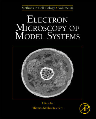
Electron Microscopy of Model Systems
Academic Press Inc (Verlag)
978-0-12-381007-6 (ISBN)
The volume covers the preparation and analysis of model systems for biological electron microscopy. The volume has chapters about prokaryotic as well as eukaryotic systems that are used as so-called model organisms in modern cell biology. These systems include the most popular systems, such as budding and fission yeast, the roundworm C. elegans, the fly Drosophila, zebrafish, mouse, and Arabidopsis, but also organisms that are less frequently used in cell biology, such as Chlamydomonas, Dictyostelium, Trypanosoma, faltworms, Axolotl and others. In addition, tissues and tissue culture systems are also covered. These systems are used for very diverse areas of cell biology, such as cell division, abscission, intracellular transport, cytoskeletal organization, tissue regeneration and others. Moreover, this issue presents the currently most important methods for the preparation of biological specimens. This volume, however, is not a classic EM methods book. The methods are not the main focus of this issue. The main goal here is to cover the methods in the context of the specific requirements of specimen preparation for each model organism or systems. This will be the first compendium covering the various aspects of sample preparation of very diverse biological systems.
Thomas Müller-Reichert is a Professor of Structural Cell Biology at the Technische Universität Dresden (TU Dresden, Germany). He is interested in how the microtubule cytoskeleton is modulated within cells to fulfill functions in mitosis, meiosis and abscission. The Müller-Reichert lab is mainly applying correlative light microscopy and electron tomography to study the 3D organization of microtubules in early embryos and meiocytes of the nematode Caenorhabditis elegans, and also in mammalian cells in culture. He has published over 75 papers and edited several volumes of the Methods in Cell Biology series on electron microscopy and CLEM. TMR obtained his PhD at the Swiss Federal Institute of Technology (ETH) in Zurich and moved afterwards for a post-doc to the EMBL in Heidelberg (Germany). He was a visiting scientist with Dr. Kent McDonald (UC Berkeley, USA). Together with Paul Verkade, he set up the electron microscope facility at the newly founded Max Planck Institute of Molecular Cell Biology and Genetics (MPI-CBG). Since 2010 he is a scientific group leader and head of the Core Facility Cellular Imaging (CFCI) of the Faculty of Medicine Carl Gustav Carus of the TU Dresden. He acted as president of the German Society for Electron Microscopy (Deutsche Gesellschaft für Elektronenmikroskopie, DGE) from 2018 to 2019. He taught numerous courses and workshops on high-pressure freezing and Correlative Light and Electron Microscopy.
Preface/Acknowledgements
Thomas Müller-Reichert
1. Electron microscopy of viruses
Michael Laue
2. Bacterial TEM: New insights from cryo-microscopy
Martin Pilhofer, Mark S. Ladinsky, Alasdair W. McDowall, and Grant Jensen*
3. Analysis of the ultrastructure of archaea by electron microscopy
Reinhard Rachel* , Carolin Meyer, Andreas Klingl, Sonja Gürster, Nadine Wasserburger, Ulf Küper, Annett Bellack, Simone Schopf, Reinhard Wirth, Harald Huber, and Gerhard Wanner
4. Chlamydomonas: Cryopreparation methods for the 3-D analysis of cellular organelles
Eileen T. O’Toole
5. Ultrastructure of the asexual blood stages of Plasmodium falciparum
Eric Hanssen* , Kenneth N Goldie, and Leann Tilley
6. Electron tomography and immunolabeling of Tetrahymena thermophila basal bodies
Thomas H. Giddings* Jr., Janet B. Meehl, Chad G. Pearson, and Mark Winey
7. Electron microscopy of Paramecium
Klaus Hausmann* and Richard D. Allen
8. Ultrastructural investigation methods for Trypanosoma brucei
Johanna L. Höög* , Eva Gluenz, Sue Vaughan, and Keith Gull
9. Dictyostelium discoideum: A model system for ultrastructural analyses of cell motility and development
Michael P. Koonce* and Ralpf Gräf
10. Towards sub-second correlative live-cell light and electron microscopy of Saccharomyces cerevisiae
Christopher Buser
11. Fission yeast: A cellular model well suited for electron microscopic investigations
Hélio D. Roque and Claude Antony*
12. High-pressure freezing and electron microscopy of Arabidopsis
Byung-Ho Kang
13. Standard and cryo-preparation of Hydra for transmission electron microscopy
Thomas W. Holstein* , Michael W. Hess, and Willi Salvenmoser
14. Electron microscopy of flatworms: Standard and cryopreparation methods
Willi Salvenmoser* , Bernhard Egger, Johannes G. Achatz, Peter Ladurner, and Michael W. Hess*
15. Three-dimensional reconstruction methods for C. elegans ultrastructure
Thomas Müller-Reichert* , Joel Mancuso, Ben Lich, and Kent McDonald*
16. Insects as model systems in cell biology
Thomas A. Keil* and R. Alexander Steinbrecht
17. Electron microscopy of amphibian model systems (Xenopus laevis and Ambystoma mexicanum)
Thomas Kurth* , Jürgen Berger, Michaela Wilsch-Bräuninger, Robert Cerny, Heinz Schwarz, Jan Löfberg, Thomas Piendl, and Hans Epperlein
18. Modern approaches for ultrastructural analysis of the zebrafish embryo
Nicole L. Schieber, Susan J. Nixon, Richard I. Webb* , and Robert G. Parton*
19. Transmission electron microscopy of cartilage and bone
Douglas R. Keene* , and Sara F. Tufa
20. Electron microscopy of the mouse central nervous system
Wiebke Möbius* , Frédérique Varoqueaux, Benjamin H. Cooper, Cordelia Imig, Torben Ruhwedel, Aiman S. Saab, and Walter Kaufmann
21. Rapidly excised and cryofixed rat tissue
Dimitri Vanhecke, Werner Graber, and Daniel Studer*
22. The actin cytoskeleton in whole mounts and sections
Guenter P. Resch* , Edit Urban, and Sonja Jacob
23. Three-dimensional cryo-electron microscopy on intermediate filaments: A critical comparison to other structural methods
Robert Kirmse, Cédric Bouchet-Marquis, Cynthia Page, and Andreas Hoenger*
24. Correlative time-lapse imaging and electron microscopy to study abscission in HeLa cells
Julien Guizetti, Jana Mäntler, Thomas Müller-Reichert* , and Daniel W. Gerlich*
25. Viral infection of cells in culture: Approaches for electron microscopy
Paul Walther* , Li Wang, Sandra Ließem, and Giada Frascaroli
26. Intracellular membrane traffic at high resolution
Jan R. T. van Weering, Edward Brown, Thomas H. Sharp, Judith Mantell, Peter J. Cullen, and Paul Verkade*
27. 3D versus 2D cell culture: Implications for electron microscopy
Michael W. Hess* , Kristian Pfaller, Hannes L. Ebner, Beate Beer, Daniel Hekl, and Thomas Seppi
28. ‘Tips and tricks’ for high-pressure freezing of model systems
Kent McDonald* , Heinz Schwarz, Thomas Müller-Reichert, Rick Webb, Christopher Buser, and Mary Morphew
| Reihe/Serie | Methods in Cell Biology ; Vol.96 |
|---|---|
| Verlagsort | San Diego |
| Sprache | englisch |
| Maße | 191 x 235 mm |
| Gewicht | 1400 g |
| Themenwelt | Naturwissenschaften ► Biologie ► Allgemeines / Lexika |
| ISBN-10 | 0-12-381007-8 / 0123810078 |
| ISBN-13 | 978-0-12-381007-6 / 9780123810076 |
| Zustand | Neuware |
| Haben Sie eine Frage zum Produkt? |
aus dem Bereich


