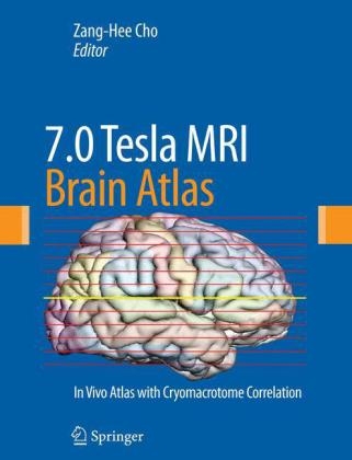
7.0 TeslaMRI Brain Atlas
In Vivo Atlas with Cryomacrotome Correlation
Seiten
2010
Humana Press Inc. (Verlag)
978-1-60761-153-0 (ISBN)
Humana Press Inc. (Verlag)
978-1-60761-153-0 (ISBN)
- Titel erscheint in neuer Auflage
- Artikel merken
Zu diesem Artikel existiert eine Nachauflage
The images in this atlas, taken at 7.0 Tesla, are of a live subject with correlating cryomacrotome photographs. They reveal details of the main stem and midbrain structures once thought impossible to visualize in vivo. Each image is annotated and detailed.
Recent advances in MRI, especially those in the area of ultra high field (UHF) MRI, have attracted significant attention in the field of brain imaging for neuroscience research, as well as for clinical applications. In "7.0 Tesla MRI Brain Atlas: In Vivo Atlas with Cryomacrotome Correlation", Zang Hee Cho and his colleagues at the Neuroscience Research Institute, Gachon University of Medicine and Science set new standards in neuro-anatomy. This unprecedented atlas presents the future of MR imaging of the brain. Taken at 7.0 Tesla, the images are of a live subject with correlating cryomacrotome photographs. Exquisitely produced in an oversized format to allow careful examination of the brain in real scale, each image is precisely annotated and detailed. The images in the Atlas reveal a wealth of details of the main stem and midbrain structures that were once thought impossible to visualize in-vivo. Ground breaking and thought provoking, "7.0 Tesla MRI Brain Atlas" is sure to provide answers and inspiration for further studies, and is a valuable resource for medical libraries, neuroradiologists and neuroscientists.
Recent advances in MRI, especially those in the area of ultra high field (UHF) MRI, have attracted significant attention in the field of brain imaging for neuroscience research, as well as for clinical applications. In "7.0 Tesla MRI Brain Atlas: In Vivo Atlas with Cryomacrotome Correlation", Zang Hee Cho and his colleagues at the Neuroscience Research Institute, Gachon University of Medicine and Science set new standards in neuro-anatomy. This unprecedented atlas presents the future of MR imaging of the brain. Taken at 7.0 Tesla, the images are of a live subject with correlating cryomacrotome photographs. Exquisitely produced in an oversized format to allow careful examination of the brain in real scale, each image is precisely annotated and detailed. The images in the Atlas reveal a wealth of details of the main stem and midbrain structures that were once thought impossible to visualize in-vivo. Ground breaking and thought provoking, "7.0 Tesla MRI Brain Atlas" is sure to provide answers and inspiration for further studies, and is a valuable resource for medical libraries, neuroradiologists and neuroscientists.
I. Axial Images of Cadaver and Human Brain of 7.0T MRI in vivo II. Sagittal Images of Cadaver and Human Brain of 7.0T MRI in vivo III. Coronal Images of Cadaver and Human Brain of 7.0T MRI in vivo
| Erscheint lt. Verlag | 26.1.2010 |
|---|---|
| Zusatzinfo | 500 colour illustrations, biography |
| Verlagsort | Totowa, NJ |
| Sprache | englisch |
| Einbandart | gebunden |
| Themenwelt | Medizinische Fachgebiete ► Chirurgie ► Neurochirurgie |
| Medizin / Pharmazie ► Medizinische Fachgebiete ► Neurologie | |
| Medizin / Pharmazie ► Medizinische Fachgebiete ► Radiologie / Bildgebende Verfahren | |
| ISBN-10 | 1-60761-153-8 / 1607611538 |
| ISBN-13 | 978-1-60761-153-0 / 9781607611530 |
| Zustand | Neuware |
| Haben Sie eine Frage zum Produkt? |
Mehr entdecken
aus dem Bereich
aus dem Bereich
Buch | Hardcover (2024)
De Gruyter (Verlag)
CHF 153,90
850 Fakten für die Zusatzbezeichnung
Buch | Softcover (2022)
Springer (Verlag)
CHF 65,80
Buch | Hardcover (2023)
Springer (Verlag)
CHF 307,95


