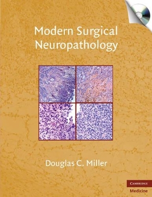
Modern Surgical Neuropathology with CD-ROM
Cambridge University Press
978-0-521-86932-4 (ISBN)
The nervous system constitutes arguably the most complex organ system in the human body, which contributes to the over-susceptibility of disease in this area. Although brain tumors were once an automatic death sentence, the outlook for recovery has brightened significantly in the past decade. The pathologist plays an indispensable role in surgical diagnosis; he or she must accurately diagnose a potentially wide array of disease entities, determine the spread of disease within millimeters, and assess patient prognosis. This new and modern reference in neuropathology comprehensively covers all the methods used by pathologists to accurately diagnose a wide array of neurologic illnesses. Brain and spinal cord tumors are the predominant focus, but a full spectrum of infectious, inflammatory, and congenital disorders are also covered in detail, in both pediatric and adult populations, with a full range of diagnostic modalities. The book is illustrated with more than 1,200 full-color photomicrographs and accompanied by a CD-ROM of all images in a downloadable format.
Douglas C. Miller was born and brought up in New York City, where he attended Stuyvesant High School. He is a graduate of Williams College (BA with highest honors in Biology, 1974) and the University of Miami School of Medicine (MD, 1978; Ph.D. in Physiology and Biophysics, 1980). Dr Miller did his residency training in Anatomic Pathology and in Neuropathology at the Massachusetts General Hospital. He has been an Assistant Professor of Pathology at the Robert Wood Johnson Medical School (1984–87), an Assistant, Associate, and then full Professor at NYU School of Medicine (1987–2007) and is now at the University of Missouri School of Medicine.
1. Introduction and overview: principles and techniques; Part I. Neoplasms; Section 1. Brain Neoplasms: Subsection 1. Intrinsic Brain Tumors: 2. Gliomas: an overview; 3. Diffuse fibrillary astrocytomas; 4. Special types of astrocytomas; 5. Oligodendroglioma; 6. Mixed gliomas; 7. Ependymomas; 8. Neuronal and neuronal-glial tumors; 9. Primitive neuroectodermal tumors; 10. Primary CNS lymphomas; 11. Primary CNS germ cell neoplasms; 12. Hemangioblastoma; 13. Choroid plexus neoplasms; 14. Metastatic neoplasms in the bran parenchyma; Subsection 2. Extrinsic Brain Tumors: 15. Meningiomas; 16. Nonmeningioma dural-based neoplasms; 17. Dura and leptominingeal metastatic cancer; 18. Tumors of intracranial peripheral nerves; 19. Craniopharyngioma and Rathke's cleft cysts; 20. Miscellaneous meningeal masses; 21. Pituitary tumors; 22. Skull-based neoplasms; Section 2. Spinal Cord Neoplasms: Subsection 1. Intrinsic Spinal Cord Tumors: 23. Intrinsic spinal cord tumors: overview; 24. Ependymomas of the spinal cord; 25. Other gliomas of the spinal cord; 26. Miscellaneous cord tumors; Subsection 2. Extrinsic Spinal Cord Neoplasms: 27. Meningiomas and other primary dural-based tumors of the spinal cord; 28. Nerve root tumors; 29. Tumors of the spine; Part II. Non-Neoplastic Mass Lesions: 30. Tumefactive demyelinating disease; 31. Inflammatory masses in the brain parenchyma; 32. Extra-axial inflammatory/infectious lesions; 33. Biopsies of cerebral infarcts; 34. Extra-axial intracranial hemorrhages; 35. Vascular malformations of the CNS parenchyma; Part III. Diagnostic Brain Biopsies for Non-Neoplastic Disease: 36. Biopsies for vasculitis; 37. Biopsies with/for non-inflammatory vascular diseases; 38. Biopsies for diagnosis of dementia; 39. Biopsies for infectious diseases; Part IV. Surgical Neuropathology of Epilepsy: 40. Current procedures leading to epilepsy surgery: what the neuropathologist should know about the epilepsy work-up prior to surgery; 41. Abnormalities presumably causing epilepsy; 42. Effects of epilepsy manifest in resected brain tissue; 43. Iatrogenic lesions; 44. Special syndromes and epilepsy.
| Zusatzinfo | 7 Tables, unspecified; 125 Plates, color; 36 Halftones, unspecified |
|---|---|
| Verlagsort | Cambridge |
| Sprache | englisch |
| Maße | 218 x 284 mm |
| Gewicht | 2340 g |
| Themenwelt | Medizinische Fachgebiete ► Chirurgie ► Neurochirurgie |
| Medizin / Pharmazie ► Medizinische Fachgebiete ► Neurologie | |
| Studium ► 2. Studienabschnitt (Klinik) ► Pathologie | |
| ISBN-10 | 0-521-86932-3 / 0521869323 |
| ISBN-13 | 978-0-521-86932-4 / 9780521869324 |
| Zustand | Neuware |
| Informationen gemäß Produktsicherheitsverordnung (GPSR) | |
| Haben Sie eine Frage zum Produkt? |
aus dem Bereich
