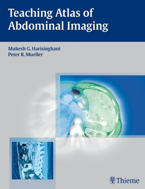
Teaching Atlas of Abdominal Imaging
Thieme Medical Publishers Inc (Verlag)
978-1-58890-656-4 (ISBN)
- Titel ist leider vergriffen;
keine Neuauflage - Artikel merken
Comprehensive review of diseases of the abdomen and pelvis
Teaching Atlas of Abdominal Imaging is a case-based reference covering the full spectrum of common and uncommon problems of the gastrointestinal and genitourinary tract encountered in everyday practice. The book organizes cases into sections based on the anatomic location of the problem. Each chapter provides succinct descriptions of clinical presentation, radiologic findings, diagnosis, and differential diagnosis for the case. The chapter then discusses the background for each diagnosis, clinical findings, common complications, etiology, imaging findings, treatment, and prognosis.
Key features:
Succinct text and consistent presentation in each chapter enhance the ease of use
Practical discussion of all current imaging modalities
Nearly 550 high-quality images demonstrate key concepts
Bulleted lists of pearls and pitfalls at the end of each chapter highlight important points
An appendix with 64-slice protocols for various CT scans, such as dual-phase liver and pancreatic scans
Ideal for both self-assessment and rapid review, this book is a valuable resource for radiologists, gastrointestinal and genitourinary radiologists, and fellows and residents in these specialties.
Associate Professor of Radiology, Director Abdominal MRI, Department of Radiology, Division of Abdominal Imaging and Interventional Radiology, Massachusetts General Hospital, Boston, MA, USA
Section 1: Liver Case 1: Hepatic Hemangioma Case 2: Focal Nodular Hyperplasia (FNH) Case 3: Liver Adenoma Case 4: Hydatid Disease Case 5: Pyogenic Liver Abscess Case 6: Hepatic Venous Malformation Case 7: Intrahepatic Cholangiocarcinoma Case 8: Hepatic Lymphoma Case 9: Liver Metastasis from Primary Pancreatic Cancer Case 10: Calcified Hepatic Metastases in a Patient with Breast Cancer Case 11: Hepatocellualr Carcinoma Section 2: Gallbladder Bile Ducts Case 12: Cholecystitis Case 13: Polyp Case 14: Porcelain Gall Bladder Case 15: Adenomyomatosis Case 16: Biliary Ascariasis Case 17: Gallbladder Carcinoma Case 18: Choledochocele Case 19: Mirizzi Syndrome Case 20: Choledocholithiasis Case 21: Primary Sclerosing Cholangitis (PSC) Case 22: Bile Leak Secondary to Bile Duct Injury Section 3: Pancreas Case 23: Pancreas Divisum Case 24: Intraductal Papillary Mucinous Neoplasia (IPMN) Case 25: Pancreatic Lymphangioma Case 26: Acute Necrotizing Pancreatitis Case 27: Pancreatic Abscess Case 28: Autoimmune Pancreatitis Case 29: Pancreatic Transection Case 30: Extramedullary Plasmacytoma Case 31: Microcystic Adenoma (serous cystadenoma) of Pancreas Case 32: Ductal Adenocarcinoma of the Pancreatic Head Case 33: Islet Cell Tumor of the Pancreas (Insulinoma) Case 34: Metastatic Renal Cell Carcinoma to the Pancreas Case 35: Solid Papillary Epithelial Neoplasm (SPEN) Case 36: Pancreatic Mucinous Cystadenocarcinoma Case 37: Primary Pacreatic Lymphoma (Non-Hodgkin s Type) Case38: Non functioning Neuroendocrine Tumor of the Pancreas Case 39: Groove Pancreatitis Case 40: Acinar Cell Carcinoma of the Pancreas (ACC) Section 4: Spleen Case 41: Accessory Spleen in Pancreatic Tail Case 42: Hemangioma of the Spleen Case 43: Splenic Sarcoidosis Case 44: Wandering Spleen Case 45: Inflammatory Splenic Pseudotumor Case 46: Autosplenectomy from Sickle Cell Disease Case 47: Thorotrast Accumation in Liver, Spleen and Lymph Nodes Case 48: Splenic Arterio-Venous Fistula with Features of Portal Hypertension. Case 49: Secondary Involvement of the Spleen in Lymphoma. Case 50: Septic Emboli Causing Infarcts in the Spleen and Left Kidney Case 51: Splenosis Section 5: Kidneys and Ureters Case 52: Autosomal Dominant Polycystic Kidney Disease (ADPKD) Case 53: Renal Angiomyolipoma Case 54: Lipid-Poor Angiomyolipoma Case 55: Atrophic Right Kidney Secondary to Reflux Nephropathy. Case 56: Renal Infarct Case 57: Pyelonephritis of the Right Kidney Case 58: Oncocytoma Case 59: Metanephric Adenoma Case 60: Renal changes from Lithium Toxicity Case 61: Localised Cystic Disease of the Kidney Case 62: Multilocular Cystic Nephroma (MLCN) Case 63: Von Hippel Lindau Disease Case 64: Cystic Renal Cell Carcinoma Case 65: Renal Abscess Case 66: Renal Cell Carcinoma (RCC) Case 67: Renal Metastases from Primary Lung Adenocarcinoma Case 68: Non-Hodgkin s Lymphoma (NHL) involving the Kidney Case 69: Retroperitoneal Fibrosis Section 6: Adrenal Glands Case 70: Adrenal Cysts Case 71: Myelolipoma Case 72: Pheochromocytoma Case 73: Adrenal Hemorrhage Case 74: Adrenal Metastases Case 75: Adrenal Carcinoma Case 76: Adrenal Ganglioneuroma Section 7: GI Tract; Peritoneal Cavity and Retroperitoneum Case 77: Abberrant Right Subclavian Artery Case 78: Gastric Mucosa-Associated Lymphoid Tissue (MALT) Lymphoma Case 79: Gastric Adenocarcinoma Case 80: Gastrointestinal Stromal Tumor (GIST) Case 81: Organoaxial Gastric Volvulus in a Large Hiatal Hernia Case 82: Duodenal Diverticulam Arising from Lateral Wall of the Duodenum Case 83: Abdominal Wall Hernia Case 84: Acute Mesenteric Ischemia Case 85: Infectious Collitis Case 86: Acute Appendicitis Case 87: Crohn Disease Case 88: Intussusception Case 89: Bowel Pneumatosis with Portal Venous Gas Case 90: Malignant Mucocele of Appendix with Pseudomyxoma Peritonei Case 91: Diverticulosis Case 92: Adenocarcinoma Sigmoid Colon with Hepatic Metastasis Case 93: Sigmoid Colonic Polyp Case 94: Ulcerative Colitis Case 95: Rectal Adenocarcinoma with Peri-Rectal Extension of Tumor Case 96: Lymphoma of the Sigmoid Colon Case 97: Acute Abdominal Attack in Familial Mediterranean Fever Case 98: Pedunculated Gastric Carcinoid, Type 3 (Sporadic subtype) Case99: Gastric Fundal Diverticulum Case 100: Gossypiboma (Textiloma or Retained Surgical Sponge) Case 101: Spigelian Hernia Case 102: Subhepatic Abscess Secondary to Dropped Gallstones Case 103: Liposarcoma Case 104: Small Bowel Carcinoid with Mesenteric Metastases Case 105: Juxtacortical Chondrosarcoma Mimicking a Pelvic Visceral Mass Case 106: Retroperitoneal Lymphangioma Case 107: Metastatic Liposarcoma with Pseudocystic Sign Case 108: Misty Mesentery Case 109: Bowel Obstruction Secondary to Oburator Hernia Case 110: Multilocular Mesothelial Inclusion Cyst Case 111: Porcelain Appendix Secondary to an Appendiceal Mucocele Case 112: Radiation Enteritis Case 113: Retroperitoneal Schwannoma Case 114: Small Bowel Obstruction Secondary to Phytobezoar Case 115: Perforated Meckel s Diverticulum Case 116: Focal Omental Infarction Case 117: Anterior Abdominal Wall Desmoid Tumor Case 118: De-differentiated Retroperitoneal Liposarcoma Case 119: Lumbar Hernia of Grynfeltt-Lesshaft Section 8: Bladder Case 120: Bladder Stone Case 121: Emphysematous Cystitis Case 122: Schisto Case 123: Hernia Case 124: Lipoma Case 125: Cancer Case 126: Urachal Adneoca Case 127: Colo-vesical fistula Section 9: Pelvis (Female Genital Pevlis) Case 128: Urethral Diverticulum Case 129: Adenomyosis Case 130: Gartner Duct Cyst Case 131: Leiomyoma Case 132: Pelvic Inflammatory Disease with Tuboovarian Abscess Case 133: Polycystic Ovary Syndrome (POS) Case 134: Gestational Trophoblastic Disease (GTD) Case 135: Endometrioma (Chocolate Cyst) Case 136: Bicornuate Uterus Case 137: Adnexal Torsion Secondary to Ovarian Dermoid Case 138: Ovarian Mucinous Cystoadenocarcinoma Case 139: Endometrial Adenocarcinoma Case 140: Cervical Carcinoma Case 141: Mature Cystic Teratoma (Dermoid Cyst0 Case 142: Cervical Lymphoma (Large B-cell, Follicular Type) (Male Genital Pevlis) Case 143: Undescended Testis (Cryptorchidism) Case 144: Segmental testicular Infarct Case 145: Left Malignant Germ Cell Tumor of the Testis with Metastatic Bulky Retroperitoneal Adenopathy Case 146: Left Extratesticular Adenomatoid Tumor Case 147: Testicular Lymphoma Case 148: Testicular Microlithiasis with Multifocal Seminoma in Left Testis Case 149: Tuberculous Epididymo-Orchitis Case 150: Prostate Carcinoma with Extracapsular and Seminal Vesicle Extension Case 151: Penile Fracture Case 152: Penile Metastases from Primary Prostate Cancer Case 153: Tubular Ectasia of the Rete Testes Case 154: Pelvic Arteriovenous Malformation (AVM) Case 155: De-differentiated Spermatic Cord Liposarcoma Section 10: Appendixes: Appendix 1: 64 Slice Protocol for Routine Abdomen and Pelvic CT Scan Appendix 2: 64 Slice Protocol for Dual Phase Liver CT Scan Appendix 3: 64 Slice CT Protocol for Evaluation of Hematuria Appendix 4: 64 Slice Protocol for Pancreatic CT Scan Appendix 5: 64 Slice CT Protocol for Evaluation of Adrenal Mass
| Verlagsort | New York |
|---|---|
| Sprache | englisch |
| Maße | 216 x 279 mm |
| Gewicht | 1678 g |
| Themenwelt | Medizinische Fachgebiete ► Innere Medizin ► Gastroenterologie |
| Medizinische Fachgebiete ► Radiologie / Bildgebende Verfahren ► Radiologie | |
| ISBN-10 | 1-58890-656-6 / 1588906566 |
| ISBN-13 | 978-1-58890-656-4 / 9781588906564 |
| Zustand | Neuware |
| Haben Sie eine Frage zum Produkt? |
aus dem Bereich


