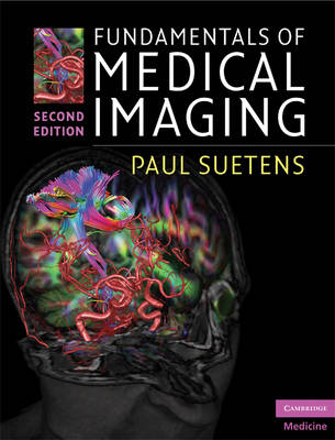
Fundamentals of Medical Imaging
Seiten
2009
|
2nd Revised edition
Cambridge University Press (Verlag)
978-0-521-51915-1 (ISBN)
Cambridge University Press (Verlag)
978-0-521-51915-1 (ISBN)
- Titel erscheint in neuer Auflage
- Artikel merken
Zu diesem Artikel existiert eine Nachauflage
An invaluable technical introduction to each imaging modality, explaining mathematical and physical principles and how medical images are obtained and interpreted. Individual chapters on each modality review the physics of the signal, image formation/reconstruction, image quality and equipment, clinical applications, biological effects and safety issues.
Fundamentals of Medical Imaging, second edition, is an invaluable technical introduction to each imaging modality, explaining the mathematical and physical principles and giving a clear understanding of how images are obtained and interpreted. Individual chapters cover each imaging modality – radiography, CT, MRI, nuclear medicine and ultrasound – reviewing the physics of the signal and its interaction with tissue, the image formation or reconstruction process, a discussion of image quality and equipment, clinical applications and biological effects and safety issues. Subsequent chapters review image analysis and visualization for diagnosis, treatment and surgery. New to this edition: • Appendix of questions and answers • New chapter on 3D image visualization • Advanced mathematical formulae in separate text boxes • Ancillary website containing 3D animations: www.cambridge.org/suetens • Full colour illustrations throughout Engineers, clinicians, mathematicians and physicists will find this an invaluable aid in understanding the physical principles of imaging and their clinical applications.
Fundamentals of Medical Imaging, second edition, is an invaluable technical introduction to each imaging modality, explaining the mathematical and physical principles and giving a clear understanding of how images are obtained and interpreted. Individual chapters cover each imaging modality – radiography, CT, MRI, nuclear medicine and ultrasound – reviewing the physics of the signal and its interaction with tissue, the image formation or reconstruction process, a discussion of image quality and equipment, clinical applications and biological effects and safety issues. Subsequent chapters review image analysis and visualization for diagnosis, treatment and surgery. New to this edition: • Appendix of questions and answers • New chapter on 3D image visualization • Advanced mathematical formulae in separate text boxes • Ancillary website containing 3D animations: www.cambridge.org/suetens • Full colour illustrations throughout Engineers, clinicians, mathematicians and physicists will find this an invaluable aid in understanding the physical principles of imaging and their clinical applications.
Paul Suetens is Professor of Medical Imaging and Image Processing, Chairman of the Medical Imaging Centre at the University Hospital Leuven, and Head of the Division for Image and Speech Processing at the Department of Electrical Engineering of K. U. Leuven, Belgium.
Preface; 1. Introduction to digital image processing; 2. Radiography; 3. X-ray computed tomography; 4. Magnetic resonance imaging; 5. Nuclear medicine imaging; 6. Ultrasound imaging; 7. Medical imaging analysis; 8. Visualization for diagnosis and therapy; Exercises; Bibliography; Index.
| Erscheint lt. Verlag | 6.8.2009 |
|---|---|
| Zusatzinfo | 76 Halftones, unspecified |
| Verlagsort | Cambridge |
| Sprache | englisch |
| Maße | 196 x 253 mm |
| Gewicht | 880 g |
| Themenwelt | Medizin / Pharmazie ► Medizinische Fachgebiete ► Radiologie / Bildgebende Verfahren |
| ISBN-10 | 0-521-51915-2 / 0521519152 |
| ISBN-13 | 978-0-521-51915-1 / 9780521519151 |
| Zustand | Neuware |
| Haben Sie eine Frage zum Produkt? |
Mehr entdecken
aus dem Bereich
aus dem Bereich
Buch | Softcover (2024)
Urban & Fischer in Elsevier (Verlag)
CHF 39,95
Buch | Softcover (2024)
John Wiley & Sons Inc (Verlag)
CHF 186,15


