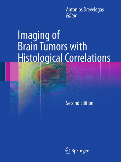
Imaging of Brain Tumors with Histological Correlations
Springer Berlin (Verlag)
978-3-540-87648-9 (ISBN)
Dr. Antonios Drevelegas is a recognized authority in the field of oncological imaging and the author of many journal publications on the subject. He currently works in the Radiology Department at the Interbalkan Medical Centre, Thessaloniki, Greece, one of Europe’s leading state-of-the-art hospitals.
Epidemiology, histologic classification and clinical course of brain tumors.- Imaging modalities in brain tumors.- Molecular abnormalities in gliomas.- Low-grade gliomas.- High-grade gliomas.- Pineal tumors.- Embryonal tumors, tumors of cranial nerves.- Meningeal tumors.- Lymphomas and hemopoietic neoplasms.- Masses of the sellar and junxtasellar region.- Brain metastasis.- Scintigraphy in brain tumors.
From the reviews:
"This book is a unique approach to linking radiographic imaging characteristics to the underlying pathology for improving the diagnosis of brain tumors. ... Extensive references are provided for the individual chapters. ... In summary, the book is very instructive and provides practical references for the diagnosis of brain tumors in relation to pathology. I do wish the book the success it deserves." (T.J. Vogl, European Radiology, Vol. 13 (6), 2003)
"This slim, well-presented book ... provides a broad overview of the imaging aspects of intracranial tumours coupled with succinct clinical and histological correlates. The layout of the book is logical and clear. ... the topics are well-covered and appropriate. The quality of the CT and MR images as well as the reproduction of the gross anatomy and histological plates is generally excellent. ... The book is ...easy to read and as such can be recommended for the general neuroradiologist." (T. Jaspan, Neuroradiology, Vol. 45 (7), 2003)
"The histologic diagnosis as well as the imaging of brain tumors are described properly, objectively, and in detail ... . This is a good book for neuroradiology fellows, but it could also be a good study tool for neuroradiologists or general radiologists working in departments with large oncologic practice ... . Of course, this book would be of interest to neurosurgeons, oncologists, and other practitioners working with brain tumors. It would be a good addition to a department library." (Sandro Rossitti, Acta Radiologica, Vol. 44 (2), 2003)
"There are clear descriptions of the techniques ... with an evaluation of their role in the primary diagnosis of brain tumours and the differentiation of tumour recurrence from radiation necrosis. ... The radiological illustrations are numerous and of good quality with a very strong emphasis on MR. ... This book is an up-to-date competent review of tumours of the cranial cavity ... . It is auseful reference book for those with a specific interest in tumours of the brain tissue itself ... ." (Dr. Evelyn Teasdale, RAD Magazine, April, 2003)
| Erscheint lt. Verlag | 22.11.2010 |
|---|---|
| Zusatzinfo | X, 432 p. |
| Verlagsort | Berlin |
| Sprache | englisch |
| Maße | 193 x 260 mm |
| Gewicht | 1335 g |
| Themenwelt | Medizin / Pharmazie ► Medizinische Fachgebiete ► Onkologie |
| Medizinische Fachgebiete ► Radiologie / Bildgebende Verfahren ► Radiologie | |
| Studium ► 2. Studienabschnitt (Klinik) ► Pathologie | |
| Schlagworte | Bildgebende Verfahren (Medizin) • Brain Tumors • CT • Hirntumor • Hirntumor / Gehirntumor • MRI • Pathology |
| ISBN-10 | 3-540-87648-0 / 3540876480 |
| ISBN-13 | 978-3-540-87648-9 / 9783540876489 |
| Zustand | Neuware |
| Informationen gemäß Produktsicherheitsverordnung (GPSR) | |
| Haben Sie eine Frage zum Produkt? |
aus dem Bereich


