Nuclear Medicine Imaging
Lippincott Williams and Wilkins (Verlag)
978-0-7817-6988-4 (ISBN)
- Titel ist leider vergriffen;
keine Neuauflage - Artikel merken
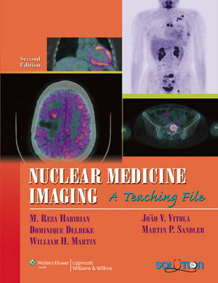
Thoroughly revised by a well-known nuclear medicine team, this teaching file reference presents 234 cases and over 600 images encompassing the gamut of procedures in contemporary clinical nuclear medicine. This Second Edition features many new cases highlighting the latest clinical and technological developments, including state-of-the-art PET/CT and SPECT/CT imaging in oncology and dramatic advances in nuclear cardiology.
Chapters present a variety of cases, from simple to complex, covering each organ system and oncologic imaging. Extensive correlative images using all relevant modalities demonstrate the use of multimodality image analysis in solving clinical problems. The final chapter focuses on common artifacts.
A companion Websitewill offer an online image bank.
SECTION HEADINGS
Chapter One Endocrine Imaging
Case 1.1 Benign Follicular Neoplasm
Case 1.2 Toxic Autonomously Functioning Adenoma
Case 1.3 Multiple Nodular Goiter with Benign Dominant Cold Nodule
Chapter Two Radionuclide Pulmonary Imaging
Case 2.1 Pulmonary Emboli with Rapid Resolution
Case 2.2 Pulmonary Emboli, Unresolved Defect
Case 2.3 High-probability Scan in Patient with Pulmonary Infiltrate
Case 2.4 Massive Pulmonary Emboli, Westermark Sign
Case 2.5 Endobronchial Mass, Mimicking PE 50
Case 2.6 Right-lower-lobe Atelectasis, Mimicking PE
Case 2.7 Intermediate-probability Scan, Triple-match Lower Lobe, Positive Angiogram
Case 2.8 Intermediate-probability Scan, Triple-match Lower Lobe, Negative Angiogram
Chapter Three Cardiovascular Imaging
Case 3.1 201Tl for MPS
Case 3.2 99mTc-labeled Agents for MPS
Case 3.3 99mTc-labeled Agents for MPS
Chapter Four Neurologic Imaging
Case 4.1 Brain Flow Present (99mTc glucoheptonate)
Case 4.2 Interictal Seizure Focus in the Right Temporal Lobe (99mTc-HMPAO)
Chapter Five Gastrointestinal and Correlative Abdominal Nuclear Medicine Imaging
Case 5.1 Gastric Emptying Study
Case 5.2 Active Small Bowel Bleeding from Postsurgical Anastomotic Site
Case 5.3 Active Gastrointestinal Bleed in the Duodenum and Sigmoid
Case 5.4 Meckel's Diverticulum
Case 5.5 Acute Gangrenous Cholecystitis
Chapter Six Renal Scintigraphy
Case 6.1 Normal Scrotal Scintigram
Case 6.2 Early Right Testicular Torsion
Case 6.3 Late Left Testicular Torsion with Acute Hemorrhage Necrosis
Case 6.4 Epididymoorchitis
Case 6.5 Right-sided Vesicoureteral Reflux
Case 6.6 Pyelonephritis
Case 6.7 Right-sided Renovascular Hypertension
Case 6.8 Bilateral Renovascular Hypertension
Chapter Seven Musculoskeletal Scintigraphy
Case 7.1 Solitary Sclerotic Metastasis
Case 7.2 Solitary Lytic Metastasis
Case 7.3 Photopenic Metastasis
Case 7.4 The Rib Lesions
Case 7.5 Progressing Metastases Versus Flare
Case 7.6 Superscan
Case 7.7 Appendicular Skeletal Metastasis
Chapter Eight Oncologic Imaging
Case 8.1 Physiologic Variation of 18F-FDG Uptake Related to Genital Tract
Case 8.2 Metastatic Melanoma and Sarcoidosis: 18F-FDG
Case 8.3 Physiological Variation 18F-FDG Uptake Related to Muscular Uptake
Case 8.4 Head and Neck Cancer: 18F-FDG PET/CT
Case 8.5 Medullary Thyroid Cancer: 18F-FDG PET/CT
Chapter Nine Artifacts
Case 9.1 Interference from a High-energy External Source
Case 9.2 Photomultiplier Tube Drift
Case 9.3 PHA Window off Peak
Case 9.4 Prosthetic Device Artifact
Case 9.5 Improper PHA Window
Case 9.6 Septal Penetration of Collimator
Case 9.7 Misadministration
Case 9.8 Center-of-rotation Error in SPECT
Index
| Erscheint lt. Verlag | 11.10.2008 |
|---|---|
| Reihe/Serie | LWW Teaching File Series |
| Verlagsort | Philadelphia |
| Sprache | englisch |
| Maße | 213 x 276 mm |
| Gewicht | 1814 g |
| Themenwelt | Medizin / Pharmazie ► Medizinische Fachgebiete ► Radiologie / Bildgebende Verfahren |
| ISBN-10 | 0-7817-6988-4 / 0781769884 |
| ISBN-13 | 978-0-7817-6988-4 / 9780781769884 |
| Zustand | Neuware |
| Haben Sie eine Frage zum Produkt? |
aus dem Bereich


