DiFiore's Atlas of Histology with Functional Correlations
Lippincott Williams and Wilkins (Verlag)
978-0-7817-7057-6 (ISBN)
- Titel erscheint in neuer Auflage
- Artikel merken
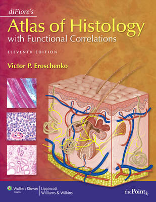
diFiore's Atlas of Histology with Functional Correlations, Eleventh Edition, explains basic histology concepts through full-color, schematic illustrations. These illustrations are supplemented by more than 450 digitized full-color online photomicrographs of histological images. Part One explains tissues and their relationship to their systems; Part Two addresses organs in a similar way. Targeting undergraduate, allied health and first and second year medical students, the 11th Edition includes new and enhanced images through redrawing and digitization to provide increased detail. This edition also features updated illustrations and information on the functions of cells, tissues, and organs of the body based on advances in research and expert recommendations. The atlas' student-friendly "Functional Correlations" sections help students study structure and function together. Students also benefit from a "realistic" perspective as more than 70 micrographs appear adjacent to color illustrations. A companion website offers student and instructor versions of diFiore's Interactive Atlas with all of the images from the book.
Introduction PART I: TISSUES Chapter 1: The Cell and the Cytoplasm Apical Surfaces of Ciliated and Nonciliated Epithelium Junctional Complex Between Epithelial Cells Basal Regions of Epithelial Cells Chapter 2: Epithelial Tissue Section 1: Classification of Epithelial Tissue Simple Squamous Epithelium: Surface View of Peritoneal Mesothelium Simple Squamous Epithelium: Peritoneal Mesothelium Surrounding Small Intestine (Transverse Section) Different Epithelial Types in the Kidney Cortex Section 2: Glandular Tissue Unbranched Simple Tubular Exocrine Glands: Intestinal Glands Simple Branched Tubular Exocrine Glands: Gastric Glands Coiled Tubular Exocrine Glands: Sweat Glands Chapter 3: Connective Tissue Loose Connective Tissue (Spread) Cells of the Connective Tissue Embryonic Connective Tissue Chapter 4: Cartilage and Bone Section 1: Cartilage Developing Fetal Hyaline Cartilage Hyaline Cartilage and Surrounding Structures: Trachea Cells and Matrix of Mature Hyaline Cartilage Section 2: Bone Endochondral Ossification: Development of a Long Bone (Panoramic View, Longitudinal Section) Endochondral Ossification: Zone of Ossification Chapter 5: Blood Human Blood Smear: Erythrocytes, Neutrophils, Eosinophils, Lymphocyte, and Platelets Human Blood Smear: Red Blood Cells, Neutrophils, Large Lymphocyte, and Platelets Erythrocytes and Platelets in Blood Smear Chapter 6: Muscle Tissue Longitudinal and Transverse Sections of Skeletal (Striated) Muscles of the Tongue Skeletal (Striated) Muscles of the Tongue (Longitudinal Section) Chapter 7: Nervous Tissue Section 1: The Central Nervous System: Brain and Spinal Cord Spinal Cord: Midthoracic Region (Transverse Section) Spinal Cord: Anterior Gray Horn, Motor Neuron, and Adjacent White Matter Spinal Cord: Midcervical Region (Transverse Section) Section 2: The Peripheral Nervous System Peripheral Nerves and Blood Vessels (Transverse Section) Myelinated Nerve Fibers (Longitudinal and Transverse Sections) Sciatic Nerve (Longitudinal Section) PART II: ORGANS Chapter 8: Circulatory System Blood and Lymphatic Vessels in the Connective Tissue Muscular Artery and Vein (Transverse Section) Chapter 9: Lymphoid System Lymph Node (Panoramic View) Lymph Node: Capsule, Cortex, and Medulla (Sectional View) Cortex and Medulla of a Lymph Node Chapter 10: Integumentary System Thin Skin: Epidermis and the Contents of the Dermis Skin: Epidermis, Dermis, and Hypodermis in the Scalp Chapter 11: Digestive System: Oral Cavity and Salivary Glands Lip (Longitudinal Section) Anterior Region of the Tongue (Longitudinal Section) Chapter 12: Digestive System: Esophagus and Stomach Wall of Upper Esophagus (Transverse Section) Upper Esophagus (Transverse Section) Chapter 13: Digestive System: Small and Large Intestines Duodenum of the Small Intestine (Longitudinal Section) Chapter 14: Digestive System: Liver, Gallbladder, and Pancreas Primate Liver Lobules (Panoramic View, Transverse Section) Chapter 15: Respiratory System Chapter 16: Urinary System Chapter 17: Endocrine System Chapter 18: Male Reproductive System Chapter 19: Female Reproductive System Chapter 20: Organs of Special Senses Index
| Erscheint lt. Verlag | 1.1.2008 |
|---|---|
| Zusatzinfo | 317 |
| Verlagsort | Philadelphia |
| Sprache | englisch |
| Maße | 216 x 280 mm |
| Gewicht | 1180 g |
| Themenwelt | Studium ► 1. Studienabschnitt (Vorklinik) ► Histologie / Embryologie |
| ISBN-10 | 0-7817-7057-2 / 0781770572 |
| ISBN-13 | 978-0-7817-7057-6 / 9780781770576 |
| Zustand | Neuware |
| Informationen gemäß Produktsicherheitsverordnung (GPSR) | |
| Haben Sie eine Frage zum Produkt? |
aus dem Bereich


