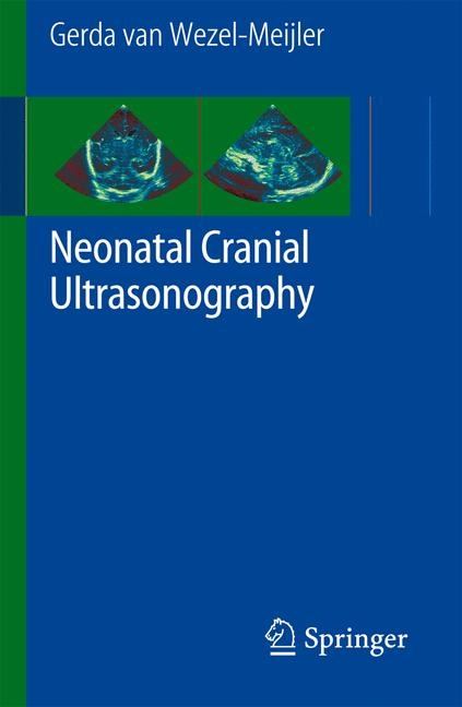
Neonatal Cranial Ultrasonography
Springer Berlin (Verlag)
978-3-540-69906-4 (ISBN)
- Titel erscheint in neuer Auflage
- Artikel merken
An exhaustive treatment of a phenomenon that causes family tragedy worldwide, this book fills a major gap in the current literature. Despite advances in neonatal care, neonatal cerebral injury remains a major cause of morbidity, mortality and disabilities. Cranial ultrasonography provides information on brain maturation in the (preterm) neonate and enables detection of frequently occurring brain anomalies in this patient group. A recent book showing high quality normal ultrasound images is lacking. This work deals with the basics of neonatal cranial ultrasonography and can be used as a reference-book providing essential information about the procedure and normal ultrasound anatomy.
Gerda Meijler is pediatrician-neonatologist. She studied medicine at the University of Amsterdam and the Academic Medical Center. She did her pediatric and neonatology training respectively at the Wilhelmina Children s Hospital, Utrecht and the Academic Medical Center, Amsterdam. After being a staff-neonatologist at the VU Medical Center, Amsterdam, where she did her PhD study on brain imaging in preterm neonates, she moved to the Leiden University Medical Center in 1999. Since then her special interest is neonatal neurology and neuro-imaging. She is the principal investigator of the Leiden neonatal neuro-imaging group. This group is actively involved in research in this field and is known for its high quality neuro-imaging (both cranial ultrasound and MRI). Together with this group Gerda has further improved neuro-imaging techniques, both for clinical and research purposes and has introduced the routine use of alternative acoustic windows in cranial ultrasonography. This has resulted in optimizing ultrasound imaging of the neonatal cerebellum. Gerda teaches neonatal neuro-imaging, both in the Netherlands and abroad at several courses and congresses. Her research now mainly focuses on imaging of the neonatal cerebellum, brain imaging and injury in the preterm neonate and on applying advanced imaging techniques in neonates. After the summer of 2011 she will move to Toronto, where she will continue her work on neonatal neurology and neuro-imaging at the neonatal units of the Hospital for Sick Children and the Mount Sinaï Hospital.
I. Theory: General introduction.- Advantages and aims of neonatal cranial ultrasonography.- Technical aspects.- Standard views, using anterior fontanel as acoustic window: coronal and sagittal planes.-Additional views using supplement acoustic windows (posterior fontanel, mastoid fontanel, temporal window).- Image assesment.- Other neuro-imaging techniques. - Maturational changes.- Summary.- Appendices. II. Ultrasound anatomy of the neonatal brain.
| Sprache | englisch |
|---|---|
| Maße | 127 x 190 mm |
| Gewicht | 285 g |
| Einbandart | Paperback |
| Themenwelt | Medizin / Pharmazie ► Medizinische Fachgebiete |
| Schlagworte | brain • Brain anatomy • Brain maturation • Cranial Ultrasonography • Hardcover, Softcover / Medizin/Klinische Fächer • HC/Medizin/Klinische Fächer • Neonate • Neugeborenes Kind • Schädel • Ultraschalldiagnostik |
| ISBN-10 | 3-540-69906-6 / 3540699066 |
| ISBN-13 | 978-3-540-69906-4 / 9783540699064 |
| Zustand | Neuware |
| Informationen gemäß Produktsicherheitsverordnung (GPSR) | |
| Haben Sie eine Frage zum Produkt? |
aus dem Bereich


