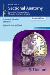
Pocket Atlas of Sectional Anatomy, Volume 1: Head and Neck
Computed Tomography and Magnetic Resonance Imaging
Seiten
2006
|
3rd edition, revised and updated
Thieme (Verlag)
978-3-13-125503-7 (ISBN)
Thieme (Verlag)
978-3-13-125503-7 (ISBN)
- Titel erscheint in neuer Auflage
- Artikel merken
Zu diesem Artikel existiert eine Nachauflage
Clear and systematic correlative guide for helping beginning and intermediate radiologists identify anatomic structures
First of three volume set which identifies the anatomical details visualized in the sectional imaging modalities
Full coverage of the head and neck!
Computed Tomography and Magnetic Resonance Imaging
Known to radiologists around the world for its superior illustrations and practical features, the Pocket Atlas of Sectional Anatomy now reflects the very latest in imaging technology. In the clinic, this compact book acts as the perfect navigational tool for radiologists and technicians working with CT and MRI.
Highlights of Volume I:
- All new CT and MR images of the highest quality, presented alongside brilliant full-color drawings
- 413 illustrations
- More slices per examination and more comprehensive coverage
- Didactic approach and consistent format throughout-one slice perpage pair
- Consistent color coding, making it easy to identify individual structuresacross several slices
Volume I: Head and Neck and its companion books
Volume II: Thorax, Heart, Abdomen, and Pelvis and
Volume III: Spine, Extremities, Joints - comprise a must-have resource for radiologists of all levels.
T. B. Moeller, E. Reif
| Reihe/Serie | Basic sciences |
|---|---|
| Pocket Atlas of Sectional Anatomy ; 1 | Thieme Flexibooks |
| Übersetzer | Adele Herzberger, Clifford Bergmann |
| Sprache | englisch |
| Gewicht | 341 g |
| Themenwelt | Medizin / Pharmazie ► Medizinische Fachgebiete |
| Schlagworte | Anatomie • Anatomie /Atlas • Atlas • Computertomographie; Atlas • Computertomographie (CT); Atlas • Hals • Kernresonanztomographie; Atlas • Kopf • Radiologie |
| ISBN-10 | 3-13-125503-X / 313125503X |
| ISBN-13 | 978-3-13-125503-7 / 9783131255037 |
| Zustand | Neuware |
| Haben Sie eine Frage zum Produkt? |
Mehr entdecken
aus dem Bereich
aus dem Bereich
Kompaktes Wissen, Sprachtraining und Simulationen für Mediziner
Buch | Softcover (2020)
Urban & Fischer in Elsevier (Verlag)
CHF 55,95


