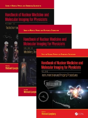
Handbook of Nuclear Medicine and Molecular Imaging for Physicists - Three Volume Set
CRC Press
978-1-032-05878-8 (ISBN)
- Titel z.Zt. nicht lieferbar
- Versandkostenfrei
- Auch auf Rechnung
- Artikel merken
This state-of-the-art set of handbooks provides medical physicists with a comprehensive overview of the field of nuclear medicine. In addition to describing the underlying, fundamental theories of the field, it includes the latest research and explores the practical procedures, equipment, and regulations that are shaping the field and it's future. This set is split into three volumes, respectively titled: Instrumentation and Imaging Procedures; Modelling, Dosimetry and Radiation Protection; and Radiopharmaceuticals and Clinical Applications.
Volume one, Instrumentation and Imaging Procedures, focuses primarily on providing a comprehensive review into the detection of radiation, beginning with an introduction to the history of nuclear medicine to the latest imaging technology. Volume two, Modelling, Dosimetry and Radiation Protection, explores the applications of mathematical modelling, dosimetry, and radiation protection in nuclear medicine. The third and final volume, Radiopharmaceuticals and Clinical Applications, highlights the production and application of radiopharmaceuticals and their role in clinical nuclear medicine practice.
These books will be an invaluable resource for libraries, institutions, and clinical and academic medical physicists searching for a complete account of what defines nuclear medicine.
The most comprehensive reference available providing a state-of-the-art overview of the field of nuclear medicine
Edited by a leader in the field, with contributions from a team of experienced medical physicists, chemists, engineers, scientists, and clinical medical personnel
Includes the latest practical research in the field, in addition to explaining fundamental theory and the field's history
Michael Ljungberg is a Professor at Medical Radiation Physics, Lund, Lund University, Sweden. He started his research in the Monte Carlo field in 1983 through a project involving a simulation of whole-body counters but later changed the focus to more general applications in nuclear medicine imaging and SPECT. As a parallel to his development of the Monte Carlo code SIMIND, he started working in 1985 with quantitative SPECT and problems related to attenuation and scatter. After obtaining his PhD in 1990, he received a research assistant position that allowed him to continue developing SIMIND for quantitative SPECT applications and establish successful collaborations with international research groups. At this time, the SIMIND program also became used world-wide. Dr. Ljungberg later became an associate professor in 1994 and he received, after a couple of years working clinically as a nuclear medicine medical physicist, a full professorship in the Science Faculty at Lund University in 2005. He became the Head of the Department of Medical Radiation Physics at Lund University in 2013 and a full professor in the Medical Faculty at Lund University in 2015. Beside from the development of SIMIND to include also new camera system such as CZT detectors, his research includes an extensive project in oncological nuclear medicine, where he, with colleagues, develop dosimetry methods based on quantitative SPECT, Monte-Carlo absorbed dose calculations, and methods for accurate 3D dose planning for internal radionuclide therapy. During the recent years, his has been focused on implementing Monte-Carlo based image reconstruction in SIMIND. He is also involved in the undergraduate education of medical physicists and bio-medical engineers and are supervising MSc and PhD students. In 2012, Professor Ljungberg became a member of the European Association of Nuclear Medicines task group on Dosimetry and served there for six years. He has published over 100 original papers, 18 conference proceedings, 18 books and book chapters and 14 peer-reviewed review papers.
Volume I: Instrumentation and Images Processing.
1. The History of Nuclear Medicine
2. Basics of Nuclear Physics
3. Basics of Radiation Interaction in Matter
4. Radionuclide Production
5. Radiometry
6. Scintillation Detectors
7. Semiconductor Detectors
8. Gamma Spectroscopy
9. Properties of the Digital Image
10. Digital Image Processing
11. Machine-Learning
12. Image File Structures in Nuclear Medicine
13. The Scintillation Camera
14. Collimators for Gamma Ray Imaging
15. Image Acquisition Protocols
16. Single Photon Emission Computed Tomography (SPECT) and SPECT/CT Hybrid Imaging
17. Dedicated Tomographic Single Photon Systems
18. Positron Emission Tomography (PET)
19. Dead Time Effects in Nuclear Medicine Imaging Studies
20. Principles of Iterative Reconstruction for Emission Tomography
21. Clinical Molecular PET/CT Hybrid Imaging
22. Clinical Molecular PET/MRI Hybrid Imaging
23. Quality Assurance of Nuclear Medicine Systems
24. Calibration and Traceability
25. Activity Quantification from Planar images
26. Quantitation in Emission Tomography
27. Multicenter studies: Hardware and Software Requirements
28. Pre-Clinical Molecular Imaging Systems
29. Monte Carlo simulations of Nuclear Medicine Imaging Systems
30. Beta and Alpha Particle Autoradiography
31. Principles behind Computed Tomography (CT)
32. Principles behind Magnetic Resonance Imaging (MRI)
Volume II: Dosimetry and Radiation Protection .
1. Introduction to Biostatistics
2. Radiobiology
3. Diagnostic Dosimetry
4. Time-Activity Curves: Data, Models, Curve Fitting and Model Selection
5. Tracer Kinetic Modelling and its use in PET Quantification
6. Principles of Radiological Protection in Healthcare
7. Controversies in Nuclear Medicine Dosimetry
8. Monte Carlo Simulation of Photon and Electron Transport in Matter
9. Patient Models for Dosimetry Applications
10. Patient-Specific Dosimetry Calculations
11. Whole Body Dosimetry
12. Personalized Dosimetry in Radioembolization
13. Thyroid Imaging and Dosimetry
14. Bone Marrow Dosimetry
15. Cellular and Multicellular Dosimetry
16. Alpha-Particle Dosimetry
17. Staff Radiation Protection
18. IAEA support to Nuclear Medicine
Volume III: Radiopharmaceuticals and Clinical Applications.
1. Principles behind Radiopharmacy
2. Radiopharmaceuticals for diagnostics: Planar/SPECT
3. Radiopharmaceuticals for diagnostics: PET
4. Radiopharmaceuticals for radionuclide therapy
5. Design Considerations for a Radiopharmaceutical Production Facility
6. Methods and Equipment for Quality Control of Radiopharmaceuticals
7. Environmental Compliance and Control for Radiopharmaceutical Production: Commercial Manufacturing and Extemporaneous Preparation
8. GMP - rules and recommendations
9. Management of Radioactive Waste in Nuclear Medicine
10. Translation of Radiopharmaceuticals: Mouse to Man
11. Radionuclide Bone Scintigraphy
12. Radionuclide Examinations of the Kidneys
13. Neuroimaging in Nuclear Medicine
14. Methodology and Clinical Implementation of Ventilation/Perfusion Tomography for Diagnosis and Follow-up of Pulmonary Embolism and Other Pulmonary Diseases Clinical use of hybrid V/P SPECT-CT
15. Myocardiac Perfusion Imaging
16. Infection and Inflammation
17. Special Considerations In Pediatric Nuclear Medicine
19. Antibody-Based Radionuclide Imaging
18. Radionuclide-Based Diagnosis and Therapy of Prostate Cancer
20. Peptide Receptor Radionuclide Therapy for Neuroendocrine Tumors
21. Lymphoscintigraphy
22. Diagnostic Ultrasound
Tomas Jansson
23. Clinical Trials - Purpose and Procedures
24. Introduction to Patient Safety and Improvement Knowledge
25. Closing remarks
| Erscheint lt. Verlag | 27.5.2024 |
|---|---|
| Reihe/Serie | Series in Medical Physics and Biomedical Engineering |
| Zusatzinfo | 128 Tables, black and white; 386 Line drawings, black and white; 287 Halftones, black and white; 673 Illustrations, black and white |
| Verlagsort | London |
| Sprache | englisch |
| Maße | 210 x 280 mm |
| Gewicht | 1700 g |
| Themenwelt | Medizinische Fachgebiete ► Radiologie / Bildgebende Verfahren ► Nuklearmedizin |
| Medizinische Fachgebiete ► Radiologie / Bildgebende Verfahren ► Radiologie | |
| Naturwissenschaften ► Physik / Astronomie ► Angewandte Physik | |
| ISBN-10 | 1-032-05878-1 / 1032058781 |
| ISBN-13 | 978-1-032-05878-8 / 9781032058788 |
| Zustand | Neuware |
| Informationen gemäß Produktsicherheitsverordnung (GPSR) | |
| Haben Sie eine Frage zum Produkt? |