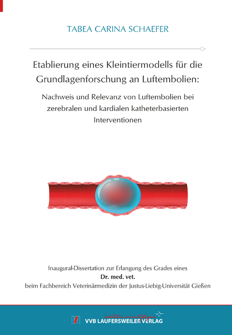
Etablierung eines Kleintiermodells für die Grundlagenforschung an Luftembolien:
Nachweis und Relevanz von Luftembolien bei zerebralen und kardialen katheterbasierten Interventionen
Seiten
2024
VVB Laufersweiler Verlag
978-3-8359-7172-1 (ISBN)
VVB Laufersweiler Verlag
978-3-8359-7172-1 (ISBN)
Iatrogene Luftembolien spielen bei unterschiedlichen zerebralen und kardialen endovaskulären Interventionen eine häufig unterschätzte Rolle für das Auftreten von postinterventionellen zerebralen Infarkten und daraus resultierenden neurologischen Funktionsstörungen. Das Gehirn ist aufgrund seiner Hypoxieempfindlichkeit und schlechten Kollateralisierung in besonderem Maße durch Luftembolien gefährdet.
In dieser Dissertation wird ein Modell zur Erforschung von Luftembolien entwickelt, mit welchem Luftblasen definierter und reproduzierbarer Größe und Anzahl produziert und im in in vivo - Modell appliziert werden können. Dieses Modell basiert auf einem Mikrofluidiksystem, mit dem die Zahl und Größe der erzeugten Luftblasen in Echtzeit unmittelbar vor der Injektion erfasst und kontrolliert werden kann. Hiermit ist eine systematische Untersuchung von Luftembolien im Kleintiermodell möglich – sowohl auf Basis von MR-tomographischen Untersuchungen in vivo als auch durch histologische Untersuchungen post mortem. Diese Methoden erlaubt, diverse Einflussfaktoren bei Luftembolien und die Pathophysiologie der daraus resultierenden Schädigungen näher zu untersuchen.
In einem ersten Schritt erfolgt die Etablierung des Modells. Es kann gezeigt werden, dass Luftblasen definierter Größe präzise und reproduzierbar erzeugt werden können (Median: 85,5 μm; SD 1,9 μm). Die automatische Zählung und Vermessung der Luftblasen erlauben eine Kontrolle der injizierten Blasen in Echtzeit. Durch die Möglichkeit, die Größe des Mikrofluidikkanals zu variieren und den Druck im System einzustellen, können Luftblasen unterschiedlicher Größe hergestellt werden. Somit bietet das Modell die Basis für systematische Studien mit unterschiedlichen Fragestellungen, etwa zum Einfluss der Bläschengröße, Bläschenzahl und des Applikationsorts auf das Infarktmuster.
In den meisten Fällen gelangt die Luft, welche Luftembolien verursacht, über einen Katheter in das Gefäßsystem. In einem zweiten Schritt werden daher Luftblasen proximal und distal eines Mikrokatheters vermessen und damit der Einfluss eines Katheters auf die Luftblasen untersucht. Es zeigt sich, dass sich nach Passage durch den Katheter der mittlere Durchmesser der Bläschen kaum verändert (Median: 86,6 μm), jedoch führt die Passage durch den Katheter zu einer größeren Streuung der Luftblasengröße (SD: 29,6 μm) und zu einer geringen Anzahl an Bläschen (60,1 % der injizierten Mikroluftblasen).
In einem dritten Schritt wird der Einfluss der Zahl der Luftblasen und des Applikationsorts auf die zerebrale Schädigung untersucht. Dabei bestätigt sich die Hypothese, dass eine höhere Anzahl an Luftblasen zu einer höheren Anzahl an Infarkten mit einem größeren totalen Infarktvolumen führt. Interessanterweise kann gezeigt werden, dass bei Injektion der gleichen Luftbläschenanzahl mit einheitlicher Blasengröße in die Aorta auf Höhe der Aortenklappe die gleiche Anzahl an zerebralen Infarkten nachzuweisen ist wie bei direkter Injektion in die A. carotis communis (5,5 vs. 5,5 (Median); p=0.769). Auch das Infarktmuster ist bei beiden Applikationsorten ähnlich. Allerdings sind die Infarkte größer, wenn die Luftblasen in die A. carotis communis injiziert werden (Infarktvolumen: 0,41 mm³ vs. 0,19 mm³ (Median); p<0.001). Dieses Ergebnis hat eine hohe klinische Relevanz, denn es lässt vermuten, dass das Risiko der Luftembolie durch kardiologische Interventionen ähnlich groß ist wie das durch neuroradiologische Interventionen. Bei Injektion in den rechten Vorhof – als Modell für die venöse Luftembolie – sind keine Infarkte nachweisbar. Dies zeigt, dass es bei der verwendeten Gesamtluftmenge im verwendeten Kleintiermodell nicht zu einer paradoxen Luftembolie kommt, am ehesten da die Filterkapazität der Lunge nicht überschritten wird.
Das im Rahmen dieser Dissertation etablierte Modell hat für weitere Untersuchungen zur Pathophysiologie, Prophylaxe und Therapie der iatrogenen Luftembolie eine hohe Relevanz, da das hier beschriebene innovative Setting (Luftblasengröße, Luftblasenzahl, Gesamtluftmenge, transfemorale Katheterisierung) weitgehend der simulierten Situation bei katheterbasierten Interventionen entspricht. Iatrogenic air embolisms play an often underestimated role in the occurrence of postinterventional cerebral infarcts, potentially resulting in neurological dysfunction, in various cerebral and cardiac endovascular interventions. The brain is particularly vulnerable to air embolisms due to its hypoxic sensitivity and poor collateralization.
In this dissertation, a model is developed to produce and inject air bubbles of defined and reproducible size and number. This model is based on a microfluidic system that allows the number and size of air bubbles to be recorded and controlled in real time immediately prior to injection. Hereby, a systematic investigation of air emboli in a small animal model is possible - by MR imaging studies in vivo as well as by histological examinations post mortem. Thus, it is possible to investigate the influencing factors in air embolism and the pathophysiology of the resulting damage in more detail.
The first step is to establish the model. It could be shown that air bubbles of defined size can be generated precisely and reproducibly (median: 85.5 μm, SD 1.9 μm). Automatic counting and measurement of the air bubble size allow real-time control of the injected bubbles. The ability to vary the size of the microfluidic channel and to adjust the pressure in the microfluidic system allows to produce air bubbles of different sizes. Thus, the model provides the basis for systematic studies on air embolisms, such as the influence of bubble size, bubble number, and application site on the infarct pattern.
In most cases of air embolisms, the air is accidentally injected into the vasculature through a catheter. Therefore, in a second step, air bubbles proximal and distal to a catheter are measured and thus the influence of a microcatheter on the air bubbles is investigated. It is found that after passage through the catheter, the mean diameter of the bubbles hardly change (median: 86.6 μm), but passage through the catheter results in a larger dispersion of the air bubble size (SD 29.6 μm) and in a lower number of air bubbles (60.1 % of the injected air bubbles).
In a third step, the influence of the number of air bubbles and the site of application on cerebral injury is investigated. This confirms the hypothesis that a higher number of air bubbles leads to a higher number of infarcts with a larger total infarct volume. Interestingly, it can be shown that when the same number of air bubbles with uniform bubble size is injected into the aorta at the level of the aortic valve, a similar number of cerebral infarcts can be detected as when the bubbles are injected directly into the carotid artery (5.5 vs. 5.5 (medians); p=0.769). Also the pattern of infarcts is comparable at both sites of application. However, infarcts are larger when air bubbles are injected into the carotid artery (median infarction volume: 0.41 mm³ vs. 0.19 mm³; p<0.001). This result has high clinical relevance as it shows that the risk of air embolism from cardiac interventions can be similar to that from neuroradiological interventions. When injected into the right atrium - as a model for venous air embolism - no infarcts are detectable. This shows that paradoxical air embolism does not occur with the total amount of air used in this model, most likely because the filtering capacity of the lung is not exceeded.
The model established in this dissertation is highly relevant for further studies on pathophysiology, prophylaxis, and therapy of iatrogenic air embolism because the innovative setting (air bubble size, air bubble number, total air volume, transfemoral catheterization) largely corresponds to the simulated situation in catheter-based interventions.
| Erscheinungsdatum | 13.02.2024 |
|---|---|
| Reihe/Serie | Edition Scientifique |
| Verlagsort | Gießen |
| Sprache | deutsch |
| Maße | 148 x 210 mm |
| Gewicht | 300 g |
| Themenwelt | Medizinische Fachgebiete ► Innere Medizin ► Pneumologie |
| Veterinärmedizin ► Allgemein | |
| Schlagworte | Embolie • Le • Verstopfung (Embolie) eines Blutgefäßes in der Lunge bzw. dem Lungenkreislauf |
| ISBN-10 | 3-8359-7172-7 / 3835971727 |
| ISBN-13 | 978-3-8359-7172-1 / 9783835971721 |
| Zustand | Neuware |
| Informationen gemäß Produktsicherheitsverordnung (GPSR) | |
| Haben Sie eine Frage zum Produkt? |
Mehr entdecken
aus dem Bereich
aus dem Bereich
Aus der Praxis für die Praxis
Buch (2022)
Thieme (Verlag)
CHF 99,40
International Trauma Life Support (ITLS)
Buch | Softcover (2024)
Hogrefe AG (Verlag)
CHF 91,00


