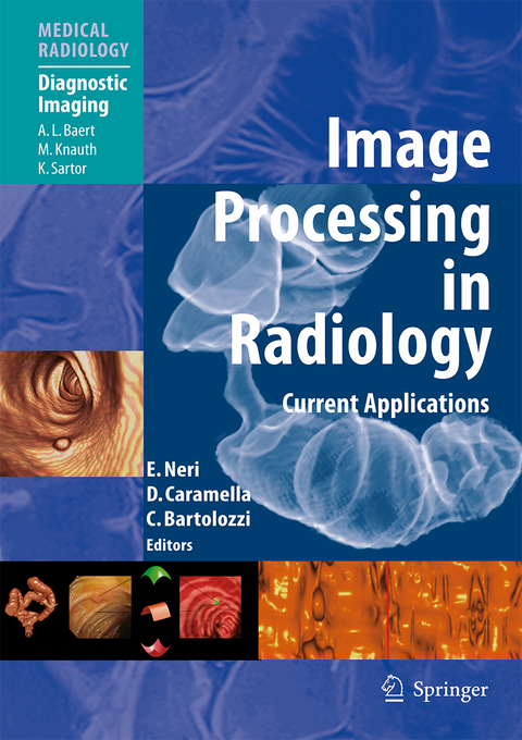
Image Processing in Radiology
Springer Berlin (Verlag)
978-3-540-25915-2 (ISBN)
Few fields have witnessed such impressive advances as image processing in radiology. The progress achieved has revolutionized diagnosis and greatly facilitated treatment selection and accurate planning of procedures. This book, written by leading experts from many countries, provides a comprehensive and up-to-date description of how to use 2D and 3D processing tools in clinical radiology. The first section covers a wide range of technical aspects in an informative way. This is followed by the main section, in which the principal clinical applications are described and discussed in depth. To complete the picture, a third section focuses on various special topics. The book will be invaluable to radiologists of any subspecialty who work with CT and MRI and would like to exploit the advantages of image processing techniques. It also addresses the needs of radiographers who cooperate with clinical radiologists and should improve their ability to generate the appropriate 2D and 3D processing.
Technical Basis of Image Processing.- US Image Acquisition.- 3D MRI Acquisition: Technique.- MDCT Image Acquisition to Enable Optimal 3D Data Evaluation.- Segmentation of Radiological Images.- Elaboration of the Images in the Spatial Domain. 2D Graphics.- 3D Medical Image Processing.- Virtual Endoscopy.- 3D Image Fusion.- Image Processing on Diagnostic Workstations.- Image Processing: Clinical Applications.- Temporal Bone.- Virtual Endoscopy of the Paranasal Sinuses.- Dental and Maxillofacial Applications.- Virtual Laryngoscopy.- Thorax.- Cardiovascular Applications.- From the Esophagus to the Small Bowel.- CT and MR Colonography.- Techniques of Virtual Dissection of the Colon Based on Spiral CT Data.- Unfolded Cube Projection of the Colon.- Liver.- Pancreas.- Biliary Tract.- Urinary Tract.- Musculoskeletal System.- Special Topics.- Clinical Applications of 3D Imaging in Emergencies.- Computer-Aided Diagnosis: Clinical Applications in the Breast.- Computer Aided Diagnosis: Clinical Applications in CT Colonography.- Ultrasound-, CT- and MR-Guided Robot-Assisted Interventions.- Virtual Liver Surgery Planning.
From the reviews:
"This comprehensive overview of advanced image processing and post-processing in diagnostic imaging covers all modalities and explains the scan parameters, fundamentals of image processing, and clinical applications. ... The book is intended primarily for practicing radiologists, although other clinicians, such as cardiologists and oncologic surgeons who use imaging extensively, may benefit as well. ... This book is very useful in explaining workstations, display functions, and advanced image processing. ... Many exam protocols are available for practicing radiologists to implement them into their routine practice." (Charles C. Matthews, Doody's Review Service, February, 2008)
"The purpose of this book is to provide extensive information on the fundamental technical aspects of present-day advanced image processing and its use in clinical applications. ... Each chapter is well written and flows well, the references are comprehensive, and the images are superb and printed on high-quality paper. ... This book belongs in the personal library and teaching libraries of radiologists, medical and surgical specialists, and any other professional dealing with advanced image processing and is an excellent book that is worth its price." (Aurelio Matamoros Jr., The Journal of Nuclear Medicine, Vol. 49, 2008)
"The book will be focused on both two-dimensional (2D) and three-dimensional (3D) aspects of the topic. ... The most likely audience for this book is radiologists without a strong technical background who are responsible for the operation of 3D image processing labs." (Bradley Erickson, Radiology, Vol. 252 (2), August, 2009)
| Erscheint lt. Verlag | 14.11.2007 |
|---|---|
| Reihe/Serie | Diagnostic Imaging | Medical Radiology |
| Vorwort | A.L. Baert |
| Zusatzinfo | X, 438 p. |
| Verlagsort | Berlin |
| Sprache | englisch |
| Maße | 193 x 270 mm |
| Gewicht | 1370 g |
| Themenwelt | Medizinische Fachgebiete ► Radiologie / Bildgebende Verfahren ► Radiologie |
| Schlagworte | 3D • 3D-Modellierung • 3D modelling • 3D-Visualisierung • 3D visualization • Bildverarbeitung • Colon • Computed tomography (CT) • Diagnosis • Endoscopy • Esophagus • Evaluation • image-guided therapy • Image Processing • Imaging • Liver • Magnetic Resonance Imaging (MRI) • Operationsvorbereitung • pancreas • Radiologie • Radiology • Surgery • Surgical Planning • Ultrasound • Virtual Reality • Volumendarstellung • volume rendering |
| ISBN-10 | 3-540-25915-5 / 3540259155 |
| ISBN-13 | 978-3-540-25915-2 / 9783540259152 |
| Zustand | Neuware |
| Haben Sie eine Frage zum Produkt? |
aus dem Bereich


