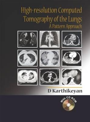
High-Resolution Computed Tomography of the Lungs: A Pattern Approach
Seiten
2005
Hodder Arnold (Verlag)
978-0-340-90580-7 (ISBN)
Hodder Arnold (Verlag)
978-0-340-90580-7 (ISBN)
- Titel ist leider vergriffen;
keine Neuauflage - Artikel merken
This highly illustrated atlas is a comprehensive yet practical guide to performing and interpreting CT imaging studies of the chest. Ideal for the clinical setting, it adopts a case-based approach for rapid reference and also includes a free companion CD of all the images from the book for teaching and/or self-assessment.
The advent of chest CT with high-resolution techniques has changed dramatically the understanding of pulmonary diseases. Conditions previously hard to distinguish using traditional film radiography, particularly the diffuse lung diseases, can now be diagnosed rapidly and the extent of the disease process identified.
Designed for easy reference in the clinical setting, this highly illustrated 'text and atlas' is a comprehensive but practical guide to performing and interpreting CT imaging studies of the chest. Opening with a review of the fundamentals of high-resolution CT in relation to lung chest anatomy, the second section forming the bulk of the book is a case-based review of both focal and diffuse lung diseases, describing the features of those diseases as visualised using CT and related differential diagnoses where relevant. The book concludes with an extensive appendix of useful information relating to chest imaging, including key facts about all the commonly encountered pathologic entities and protocols that can be referred to in the clinical setting.
The book is accompanied by a CD containing all the images from the book with a presentation on pattern approach that can be used for teaching purposes as well as self-assement.
D. Karthikeyan, Chief Radiologist, Division of Computed Tomography and Body Imaging, Department of Imaging Sciences, KG Hospital and Postgraduate Institute; and Programme Director, Radeducation Pvt. Ltd, Coimbatore, India.
| Erscheint lt. Verlag | 29.7.2005 |
|---|---|
| Zusatzinfo | 13 b/w line drawings, 188 b/w halftones, 6 col. line drawings, 5 col. halftones |
| Verlagsort | London |
| Sprache | englisch |
| Maße | 210 x 280 mm |
| Themenwelt | Medizinische Fachgebiete ► Innere Medizin ► Pneumologie |
| Medizinische Fachgebiete ► Radiologie / Bildgebende Verfahren ► Computertomographie | |
| ISBN-10 | 0-340-90580-8 / 0340905808 |
| ISBN-13 | 978-0-340-90580-7 / 9780340905807 |
| Zustand | Neuware |
| Haben Sie eine Frage zum Produkt? |
Mehr entdecken
aus dem Bereich
aus dem Bereich
Aus der Praxis für die Praxis
Buch (2022)
Thieme (Verlag)
CHF 99,40
International Trauma Life Support (ITLS)
Buch | Softcover (2024)
Hogrefe AG (Verlag)
CHF 91,00


