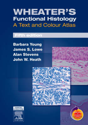
Wheater's Functional Histology
A Text and Colour Atlas
Seiten
2006
|
5th Revised edition
Churchill Livingstone (Verlag)
978-0-443-06850-8 (ISBN)
Churchill Livingstone (Verlag)
978-0-443-06850-8 (ISBN)
- Titel erscheint in neuer Auflage
- Artikel merken
Zu diesem Artikel existiert eine Nachauflage
Contains over 900 images and illustrations to help you learn and review the microstructure of human tissues. This atlas includes a section on general cell structure and replication. It covers basic tissue types and presents the microstructures of each of the major body systems.
This title is highly commended, Basic and Clinical Sciences Category, BMA Awards 2007! This best-selling atlas contains over 900 images and illustrations to help you learn and review the microstructure of human tissues. It starts with a section on general cell structure and replication. Basic tissue types are covered in the following section, and the third section presents the microstructures of each of the major body systems. The highest-quality color light micrographs and electron micrograph images are accompanied by concise text and captions which explain the appearance, function, and clinical significance of each image. The accompanying website lets you view all the images from the atlas with a "virtual microscope", allowing you to view the image at a variety of pre-set magnifications.
This title is highly commended, Basic and Clinical Sciences Category, BMA Awards 2007! This best-selling atlas contains over 900 images and illustrations to help you learn and review the microstructure of human tissues. It starts with a section on general cell structure and replication. Basic tissue types are covered in the following section, and the third section presents the microstructures of each of the major body systems. The highest-quality color light micrographs and electron micrograph images are accompanied by concise text and captions which explain the appearance, function, and clinical significance of each image. The accompanying website lets you view all the images from the atlas with a "virtual microscope", allowing you to view the image at a variety of pre-set magnifications.
Part 1 - The Cell 1.Cell structure and function 2.Cell cycle and repetition Part 2 - Basic Tissue Types 3.Blood 4.Supporting/connective tissues 5.Epithelial tissues 6.Muscle 7.Nervous tissues Part 3 - Organ systems 8.Circulatory system 9.Skin 10.Skeletal tissues 11.Immune system 12.Respiratory system 13.Oral tissues 14.Gastrointestinal tract 15.Liver and pancreas 16.Urinary system 17.The endocrine glands 18.Male reproductive system 19.Female reproductive system 20.Central nervous system 21.Special sense organs
| Erscheint lt. Verlag | 14.3.2006 |
|---|---|
| Zusatzinfo | Approx. 850 illustrations (700 in full color) |
| Verlagsort | London |
| Sprache | englisch |
| Maße | 210 x 297 mm |
| Themenwelt | Medizin / Pharmazie ► Medizinische Fachgebiete |
| Studium ► 1. Studienabschnitt (Vorklinik) ► Histologie / Embryologie | |
| Schlagworte | Histologie; Atlas |
| ISBN-10 | 0-443-06850-X / 044306850X |
| ISBN-13 | 978-0-443-06850-8 / 9780443068508 |
| Zustand | Neuware |
| Haben Sie eine Frage zum Produkt? |
Mehr entdecken
aus dem Bereich
aus dem Bereich
Zytologie, Histologie und mikroskopische Anatomie
Buch | Hardcover (2022)
Urban & Fischer in Elsevier (Verlag)
CHF 75,60
Gewebelehre, Organlehre
Buch | Spiralbindung (2024)
Urban & Fischer in Elsevier (Verlag)
CHF 34,95


