Artificial Intelligence in PET/CT Oncologic Imaging
Springer International Publishing (Verlag)
978-3-031-10092-5 (ISBN)
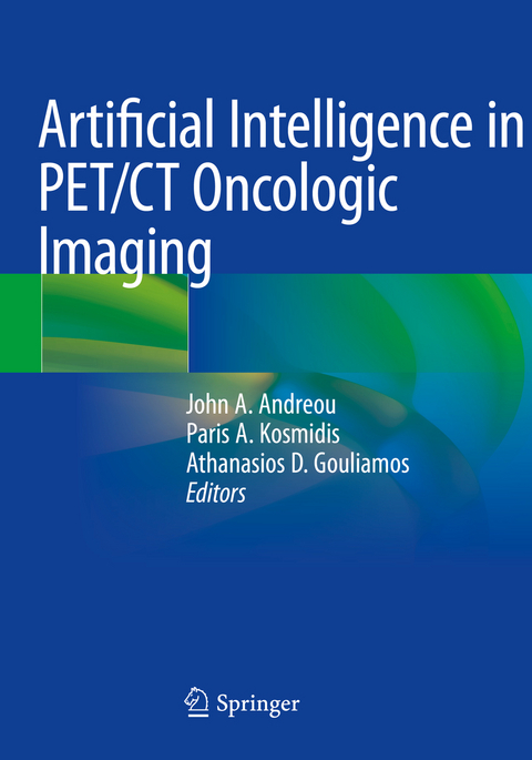
Originally positioned as a means for noninvasive molecular phenotyping and quantification in the 1970s, PET's technological improvements in the 2000s generated renewed interest in quantification, which has grown over the last five years. This progress is parallel with the development of Artificial intelligence (AI) systems for Oncology which aim at providing the best possible treatment to patients suffering from lung, breast, brain, prostate, liver and other types of cancer.
The chapters provide an overview of the use of AI in PET/CT imaging for various types of cancer, and it will be an invaluable tool especially for nuclear medicine physicians and oncologists.
lt;p>John A. Andreou is Chairman of the Imaging Departments at Hygeia and Mitera Hospitals, Greece. He got his PhD at the Athens University Medical School in 1983. Then, he worked as Adjunct Assistant Professor of Radiology at the same university. In 1984 he was Director of the Section of Computed Tomography at the Agios Savvas Cancer Hospital. From 1986 to 2011 he was Director of the Section of Computed Tomography and Magnetic Resonance at Hygeia Hospital as well as Director of the PET/CT section from 2004 to 2011. Prof. Andreou authored more than 50 original papers, reviews or book chapters in Greek and international journals/medical textbooks. He is co-editor of the books: "Imaging in Clinical Oncology" (Springer, 2014 and 2nd ed 2018) and "PET/CT in Lymphomas: a case based atlas" (Springer, 2016). He did more than 130 presentations at Greek and International medical conferences and participated in more than 170 lectures, seminars, postgraduate courses and round tables, including chairing many of these.
Paris A. Kosmidis is Head of the second Medical Oncology Department at "HYGEIA" Hospital in Athens, Greece and former ESMO President. He received appointments to serve as Head of the Second Department of Medical Oncology at St. Anargiri Cancer Hospital in Athens (1981 - 1985) and at "METAXA" Cancer Hospital in Piraeus (1985 - 1997). He published more than one hundred papers and completed 5 books. He serves at the Editorial Board in many oncology journals of the world. Dr Kosmidis has been awarded by the Academy of Athens twice and he is honorary member of ESMO. He is co-editor of the books: "Imaging in Clinical Oncology" (Springer, 2014 and 2nd ed 2018) and "PET/CT in Lymphomas: a case based atlas" (Springer, 2016).
Athanasios D. Gouliamos is Professor of Radiology, Emeritus, at the National and Kapodistrian University of Athens Medical School. He has previously worked as Chairman of the 1st Radiology Department at Aretaieion Hospital (2008-2013) and the 2nd Radiology Department at Attikon Hospital, University of Athens Medical School (2005-2008). He also served as co-director of the 12th and 13th Cycle of ESNR-ECNR Pierre Lasjaunias Courses in Neuroradiology, Diagnostic and Interventional (2013-2016). His research interest is primarily in the fields of Neuroradiology (Stroke), Advanced MRI Techniques in Medicine and Biomaterials and Radiology Information & Forecasting Integrated Systems. He authored more than 170 Pubmed-cited publications and is also author and co-author of books for medical students, published in Greece. He is co-editor of the books: "Imaging in Clinical Oncology" (Springer, 2014 and 2nd ed 2018) and "PET/CT in Lymphomas: a case based atlas" (Springer, 2016). He served as reviewer in European Radiology and has been awarded from the Athens Academy as co-author of the book Introduction in Medical Imaging (1992).
1. Introduction : Artificial intelligence (AI) systems for Oncology.- 2. PET in Bone and Soft tissue tumors.- 3. (CNS) PET/CT: current AI applications.- 4. PET/CT findings in Head and Neck Cancer, current AI applications.- 5. PET/CT in Lung cancer, current AI applications.- 6. Breast cancer: PET/CT imaging, AI applications.- 7. PET/CT in Gynecologic cancer, current AI applications.- 8. PET/CT in Rectal cancer, current AI applications.- 9. PET/CT in Neuroendocrine tumors, current AI applications.- 10. PET/CT in the evaluation of Adrenal gland mass.- 11. PET/CT in Renal cancer.- 12. PET/CT in Testicular cancer.- 13. PET/CT in Prostate cancer.- 14. PET/CT in Malignant Lymphomas.
| Erscheinungsdatum | 24.10.2023 |
|---|---|
| Zusatzinfo | XIV, 151 p. 27 illus., 24 illus. in color. |
| Verlagsort | Cham |
| Sprache | englisch |
| Maße | 178 x 254 mm |
| Gewicht | 330 g |
| Themenwelt | Medizin / Pharmazie ► Medizinische Fachgebiete ► Radiologie / Bildgebende Verfahren |
| Schlagworte | future of medicine • machine learning • Neural networks • NSCSL • Personalized medicine • Radiomics |
| ISBN-10 | 3-031-10092-1 / 3031100921 |
| ISBN-13 | 978-3-031-10092-5 / 9783031100925 |
| Zustand | Neuware |
| Haben Sie eine Frage zum Produkt? |
aus dem Bereich


