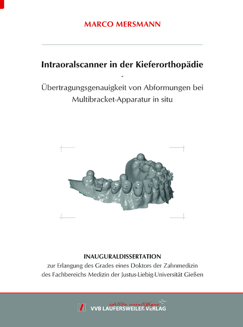
Intraoralscanner in der Kieferorthopädie - Übertragungsgenauigkeit von Abformungen bei Multibracket-Apparatur in situ
Seiten
2023
VVB Laufersweiler Verlag
978-3-8359-7121-9 (ISBN)
VVB Laufersweiler Verlag
978-3-8359-7121-9 (ISBN)
- Keine Verlagsinformationen verfügbar
- Artikel merken
Das Vorhandensein einer Multibracket-Apparatur hat unterschiedliche Auswirkungen auf die Ergebnisse einer Abformung und beeinflusst infolgedessen entscheidend die Qualität der kieferorthopädischen Behandlung. Ziel dieser In-vitro-Untersuchung war es, die Übertragungsgenauigkeit von Intraoralscannern bei Vorliegen einer festsitzen-den Multibracket-Apparatur zu untersuchen. Hierbei wurde ein Vergleich zwischen der konventionellen Alginatabformung mit anschließender Modellherstellung mit Su-perhartgips Typ IV und der digitalen Modellherstellung durch fünf Intraoralscanner (CS 3600, Carestream Dental, Rochester, NY, USA; Primescan, Dentsply Sirona, Bensheim, Deutschland; Trios 4, 3Shape, Kopenhagen, Dänemark; i500, Medit, Se-oul, Südkorea; Emerald S, Planmeca, Helsinki, Finnland) durchgeführt.
Vier Referenzpunkte in Form von Metallkugeln wurden vor der Alginatabformung und der Digitalisierung durch die Intraoralscanner auf das eigens für die vorgenom-menen Versuche hergestellte Unterkiefermodell platziert. Die starre Positionierungs-platte aus Metall, in welcher die Kugeln magnetisch gesichert wurden, ermöglichte eine reproduzierbare Platzierung der Kugeln und diente zudem als ergebnissichernde Referenz für die Kugelpositionen. Im ersten Studiendurchgang wurde das Unterkie-fermodell ohne Brackets, im zweiten und im dritten Studiendurchgang mit Metallbra-ckets sowie im vierten und im fünften Studiendurchgang mit Keramikbrackets verse-hen und jeweils digitalisiert. Ergänzend wurden die Studiendurchgänge drei und fünf mit einem an den Brackets befestigten metallischen Bogen durchgeführt. Die einzel-nen Arbeitsschritte der jeweiligen Abformung wurden zeitlich erfasst, tabellarisch protokolliert und im Folgenden gegenübergestellt. In Summe konnten nach fünf Stu-diendurchgängen 300 (n = 12x5x5) digitalisierte Modelle mit einer 3D-Software (GOM Inspect, Braunschweig, Deutschland) analysiert und 60 (n = 12x5) Gipsmodel-le mit einem Koordinatenmessgerät ausgewertet werden. Die gemessenen linearen Distanzen und Winkel zwischen den Kugelmittelpunkten konnten infolge mit Refe-renzdaten verglichen werden.
Das Resultat der Auswertung und der statistischen Analyse der Distanz- und Win-kelmessungen und der Zeiterfassung waren signifikante Unterschiede (p < 0,05) in-nerhalb der Studiendurchgänge zwischen den verschiedenen Abformmethoden. Die Intraoralscanner zeigten bei Vorliegen einer Multibracket-Apparatur ohne sowie mit Bogen eine höhere Übertragungsgenauigkeit im Vergleich zum herkömmlichen Ar-beitsablauf Alginatabformung und konventionelle Herstellung von Gipsmodellen.
Aufgrund dieser In-vitro-Studie können Intraoralscanner für eine Ganzkieferabfor-mung, bei Vorliegen einer Multibracket-Apparatur ohne sowie auch mit einem metal-lischen Bogen, für den kieferorthopädischen Alltag empfohlen werden. Ohne das Vorhandensein einer Multibracket-Apparatur in situ kann mit der konventionellen Methode – Alginatabformung und Modellherstellung – eine höhere Genauigkeit er-zielt werden. The presence of a multibracket appliance has different effects on the results of an impression and consequently has a decisive influence on the quality of orthodontic treatment. The aim of this in vitro study was to investigate the transfer accuracy of intraoral scanners in the presence of a fixed multibracket appliance. A comparison was made between conventional alginate impression taking followed by model fabri-cation with superhard stone type IV and digital model fabrication using five intraoral scanners (CS 3600, Carestream Dental, Rochester, NY, USA; Primescan, Dentsply Sirona, Bensheim, Germany; Trios 4, 3Shape, Copenhagen, Denmark; i500, Medit, Seoul, South Korea; Emerald S, Planmeca, Helsinki, Finland).
Four reference points in the form of metal spheres were placed on the mandibular model (made specifically for the tests performed) before alginate impressions were taken and digitized by the intraoral scanners. The rigid metal positioning plate, in which the spheres were magnetically secured, allowed reproducible placement of the spheres and also served as a result-securing reference for the sphere positions. In the first study cycle, the mandibular model was provided without brackets, in the second and third study cycles with metal brackets and in the fourth and fifth study cycles with ceramic brackets and digitized in each case. In addition, study cycles three and five were performed with a metal wire attached to the brackets. The individual steps of impression taking were recorded in time, logged in tabular form and compared in the following. In total, 300 (n = 12x5x5) digitized models were analyzed with 3D software (GOM Inspect, Braunschweig, Germany) and 60 (n = 12x5) plaster models were evaluated with a coordinate measuring machine after five study cycles. As a result, the measured linear distances and angles between the sphere centers could be compared with reference data.
The result of the evaluation and the statistical analysis of the distance and angle measurements and the time recording show significant differences (p < 0.05) within the study cycles between the different impression methods. The intraoral scanners showed higher transfer accuracy in the presence of a multibracket appliance without as well as with wire compared to the conventional workflow of alginate impression taking and conventional fabrication of plaster models.
Based on this in vitro study, intraoral scanners for full-arch impression taking in the presence of a multibracket appliance without as well as with a metallic wire can be recommended for everyday orthodontic use. Without the presence of a multibracket appliance in situ, higher accuracy can be achieved with the conventional method – alginate impression taking and model fabrication.
| Erscheinungsdatum | 02.06.2023 |
|---|---|
| Reihe/Serie | Edition Scientifique |
| Verlagsort | Gießen |
| Sprache | deutsch |
| Maße | 148 x 215 mm |
| Gewicht | 460 g |
| Themenwelt | Medizin / Pharmazie ► Zahnmedizin ► Klinik und Praxis |
| Medizin / Pharmazie ► Zahnmedizin ► Kieferorthopädie | |
| Schlagworte | CAD • CAM • Kiefer • Zahn |
| ISBN-10 | 3-8359-7121-2 / 3835971212 |
| ISBN-13 | 978-3-8359-7121-9 / 9783835971219 |
| Zustand | Neuware |
| Informationen gemäß Produktsicherheitsverordnung (GPSR) | |
| Haben Sie eine Frage zum Produkt? |
Mehr entdecken
aus dem Bereich
aus dem Bereich
Prüfungswissen Kariologie, Endodontologie und Parodontologie
Buch (2023)
Deutscher Ärzteverlag
CHF 97,95


