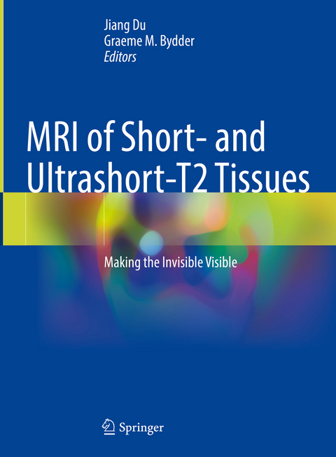
MRI of Short- and Ultrashort-T2 Tissues
Springer International Publishing (Verlag)
978-3-031-35196-9 (ISBN)
This is an ideal guide for physicists and radiologists interested in learning more about the use of UTE and ZTE type techniques for MRI of short and ultrashort-T2 tissues.
lt;p>Dr Jiang Du is a physicist who is Professor of Radiology and Director of the Ultrashort Echo Time (UTE) Imaging Lab at the University of California, San Diego (UCSD). His research focuses on the development of magnetic resonance imaging (MRI), especially UTE MRI of short and ultrashort-T2 tissues such as cortical bone, tendons, ligaments, menisci, iron containing tissues and myelin. He has published over 200 papers and mentored more than 100 trainees. Currently,he is a principal investigator for three NIH RO1 grants and one VA merit grant to study osteoarthritis (OA), osteoporosis (OP), Alzheimer's disease (AD), and traumatic brain injury (TBI). He has served on many academic organizations and review committees, including study sections for the National Institute of Health (NIH), Department of Defense (DOD), and Radiological Society of North America (RSNA). He has won numerous awards including the RSNA Research Scholarship, the American Heart Association (AHA) Young Investigator Award, the International Society of Magnetic Resonance in Medicine (ISMRM) Outstanding Teacher Award, the International Skeletal Society (ISS) Excellence Award, and the Academy for Radiology & Biomedical Imaging Research (ARBIR) Distinguished Investigator Award. Dr Du is Fellow of the ISMRM and American Institute for Medical and Biological Engineering (AIMBE).
Dr Graeme Bydder is a radiologist who began clinical MRI in 1981 and has continued to the present day. He was a joint author of the first paper on the use of MRI in multiple sclerosis in 1981 and the first comprehensive paper on MRI of the brain in 1982. He was also a joint author of many of the first papers describing techniques now commonly used in clinical imaging. These include: inversion recovery and heavily T2-weighted spin echo sequences, Gd based contrast agents, STIR, susceptibility weighted imaging and FLAIR. He has worked at UCSD since 2003 on UTE development which uses radial imaging and high performance gradients to detect signals from bone, tendon, ligaments and other short and ultrashort-T2 tissues. He is also working on multiplied, added, subtracted and divided inversion recovery (MASDIR) sequences which provide improved lesion contrast in clinical MRI. He is a past president of the ISMRM, gold medalist of the ISMRM and Royal College of Radiologists, as well as an honorary member of the American and British Societies of Neuroradiology.Part I: UTE MRI - Data Acquisition.- Basic Principles of MRI.- An Introduction to UTE MRI.- 2D UTE MRI.- 3D UTE MRI.- ZTE MRI.- PETRA MRI.- SWIFT MRI.- WASPI MRI.- Hybrid 3D UTE (Stack of STAR, AWSOS).- Cartesian Variable TE MRI.- Part II: UTE MRI - Contrast Mechanisms.- UTE with Echo Subtraction.- UTE with on/off Resonance Saturation.- UTE with Adiabatic Inversion (Single IR, Dual IR, Double IR, IR Fat Sat, DESIRE, STAIR).- UTE with Water Excitation.- UTE Fat/Water Imaging (UTE IDEAL, UTE Single Point Imaging).- UTE Spectroscopic Imaging.- Pulse Sequence as Tissue Property Filters.- Clinical Use of MASTIR Pulse Sequences.- Part III: UTE MRI - Quantification.- UTE T1 Quantification.- UTE T2* Quantification.- UTE Looping Star T2* Quantification.- UTE T1 Quantification.- UTE Proton Density Quantification.- UTE Magnetization Transfer Imaging.- UTE Quantitative Susceptibility Mapping.- UTE Perfusion.- UTE Diffusion.- UTE with deep learning for fully automated segmentation and quantitativemapping.- Part IV: UTE MRI - Applications.- UTE T2* in Osteoarthritis.- UTE MRI Biomarker Panel in Osteoarthritis.- UTE Porosity Index and Suppression Ratio in Osteoporosis.- UTE Bound Water and Pore Water in Osteoporosis.- UTE MRI Biomarker Panel in Osteoporosis.- UTE MRI in the Spine.- UTE MRI in Tendinopathy.- UTE MRI in Psoriatic Arthropathy.- UTE MRI in Hemophilia Arthropathy.- UTE MRI in Temporomandibular Disorders.- UTE MRI in Multiple Sclerosis.- UTE MRI in Traumatic Brain Injury.- UTE MRI in the Lung.- UTE MRI in the Liver.- UTE MRI in cerebral aneurysm and coil embolization.- UTE MRI in Vascular Calcification.- UTE MRI in Cryotherapy.- UTE MRI of iron nanoparticles.- UTE in PET/MRI.- UTE MRI in "CT-like" bone imaging.- Challenges and future directions in UTE imaging.
| Erscheinungsdatum | 23.02.2024 |
|---|---|
| Zusatzinfo | XXVII, 611 p. 414 illus., 302 illus. in color. |
| Verlagsort | Cham |
| Sprache | englisch |
| Maße | 210 x 279 mm |
| Gewicht | 2092 g |
| Themenwelt | Medizin / Pharmazie ► Medizinische Fachgebiete ► Radiologie / Bildgebende Verfahren |
| Schlagworte | contrast mechanism • MRI • Quantitative • short-T2 tissues • Ute |
| ISBN-10 | 3-031-35196-7 / 3031351967 |
| ISBN-13 | 978-3-031-35196-9 / 9783031351969 |
| Zustand | Neuware |
| Informationen gemäß Produktsicherheitsverordnung (GPSR) | |
| Haben Sie eine Frage zum Produkt? |
aus dem Bereich


