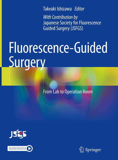
Fluorescence-Guided Surgery
Springer Verlag, Singapore
978-981-19-7371-0 (ISBN)
Fluorescence-Guided Surgery – From Lab to Operation Room is recommended for surgeons, operating nurses, medical experts, basic researchers and, industry engineers worldwide beyond boundaries of specialties. Edited and written by experts of The Japanese Society for Fluorescence-Guided Surgery, those who are the founders of the technology, it describes the accurate development history and cutting-edge techniques based on the knowledge accumulated over the years.
Takeaki Ishizawa, MD, PhD, FACSDepartment of Hepatobiliary-Pancreatic Surgery,Graduate School of Medicine, Osaka Metropolitan UniversityOsakaJapan Editorial Supervisor: Japanese Society for Fluorescence Guided Surgery (JSFGS)
Part I Basics of intraoperative fluorescence imaging [Introduction].- Chapter 1 Clinically available fluorescent reagents.- Chapter 2 ICG fluorescence imaging system for open surgery.- Chapter 3 ICG fluorescence imaging system for endoscopic surgery and robot-assisted surgery.- Chapter 4 5-ALA fluorescence imaging system.- Chapter 5 How to introduce fluorescence imaging to the operating room.- Chapter 6 Recording of intraoperative fluorescence imaging.- Part II Intraoperative Fluorescence Imaging [Practice] - Perfusion assessment.- Chapter 7 Introduction.- Chapter 8 Coronary angiography.- Chapter 9 Cerebral angiography (cerebral aneurysm).- Chapter 10 Evaluation of blood perfusion in skin flaps.- Chapter 11 Evaluation of blood perfusion in the upper gastrointestinal tract.- Chapter 12 Evaluation of blood perfusion in colorectal surgery.- Chapter 13 Perfusion assessment in HBP surgery and liver transplantation.- Part III Intraoperative Fluorescence Imaging [Practice] - Imaging of cancer.- Chapter 14 Liver cancer (primary liver cancer, metastatic liver cancer).- Chapter 15 Lung cancer (marking the tumor site).- Chapter 16 Gastric cancer (primary tumor, peritoneal dissemination).- Chapter 17 Brain tumor.- Chapter 18 Bladder cancer.- Part IV Intraoperative Fluorescence Imaging [Practice] - Imaging of lymph nodes and lymph vessels.- Chapter 19 Introduction.- Chapter 20 Identification of sentinel lymph nodes in breast cancer surgery.- Chapter 21 Identification of sentinel lymph nodes in gastric cancer surgery.- Chapter 22:Identification of sentinel lymph nodes in colorectal cancer surgery.- Chapter 23 Identification of sentinel lymph nodes in gynecologic surgery.- Chapter 24 Lymphography and evaluation of lymphedema.- Part V Intraoperative Fluorescence Imaging [Practice] - Imaging of anatomical structures.- Chapter 25 Imaging of the bile ducts (fluorescence cholangiography).- Chapter 26 Hepatic segmentation.- Chapter 27 Lung segmentation.- Chapter 28 Visualization of the ureter.- Chapter 29 Imaging of the parathyroid gland.- Part VI Intraoperative Fluorescence Imaging in Practice [Development].- Chapter 30 Development of novel fluorescent probes - Rapid Intraoperative Visualization of Microcarcinoma by Local Application of Chemical Fluorescence Probes.- Chapter 31 Development of a new imaging system.- Chapter 32 Development of a new operating room that integrates imaging information.- Chapter 33 Therapeutic applications (1): Photodynamic therapy using porphyrin compounds.- Chapter 34 Application to therapy (2): Photoimmunotherapy using a near-infrared fluorescent probe.
| Erscheinungsdatum | 30.11.2023 |
|---|---|
| Co-Autor | JSFGS |
| Zusatzinfo | 177 Illustrations, color; 7 Illustrations, black and white; XIX, 263 p. 184 illus., 177 illus. in color. |
| Verlagsort | Singapore |
| Sprache | englisch |
| Original-Titel | Jutsuchu Keikou Imaging Jissen Guide – Labo kara Opeshitu made- |
| Maße | 210 x 279 mm |
| Themenwelt | Medizin / Pharmazie ► Medizinische Fachgebiete ► Chirurgie |
| Medizin / Pharmazie ► Medizinische Fachgebiete ► Onkologie | |
| Studium ► 1. Studienabschnitt (Vorklinik) ► Anatomie / Neuroanatomie | |
| Schlagworte | Fluorescence-guided surgery • Intraoperative fluorescence imaging • Near-infrared imaging • photodynamic therapy • Photoimmunotherapy • Theranostics |
| ISBN-10 | 981-19-7371-7 / 9811973717 |
| ISBN-13 | 978-981-19-7371-0 / 9789811973710 |
| Zustand | Neuware |
| Informationen gemäß Produktsicherheitsverordnung (GPSR) | |
| Haben Sie eine Frage zum Produkt? |
aus dem Bereich


