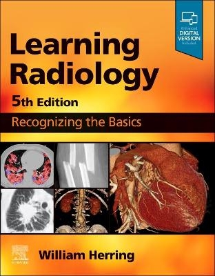
Learning Radiology
Elsevier - Health Sciences Division (Verlag)
978-0-323-87817-3 (ISBN)
Uses a clear, conversational writing style-with a touch of humor-to explain what you need to know to effectively interpret medical images of all modalities.
Teaches how to arrive at a diagnosis by following a pattern recognition approach, and logically overcome difficult diagnostic challenges with the aid of decision trees.
Employs an easy-to-read, bullet-point format, including bolded key points and icons designating special content: Diagnostic Pitfalls, Really Important Points, Take-Home Points, and Weblinks.
Features more than 850 high-quality illustrations, useful tables, case study questions, and teaching boxes throughout.
Shares the extensive knowledge and experience of esteemed author Dr. William Herring, a skilled radiology teacher and the host of his own specialty website, www.learningradiology.com.
Offers quick review and instruction for medical students, residents, and fellows, as well as those in related fields such as nurse practitioners and physician assistants.
An eBook version is included with purchase. The eBook allows you to access all of the text, figures and references, with the ability to search, customize your content, make notes and highlights, and have content read aloud-as well as access bonus content, including new appendices covering the Discovery of X-rays, Diagnostic Radiology Signs, and Artificial Intelligence in Radiology; USMLE-style Q&A; 30 videos; and more.
1. Recognizing Anything: Past, Present and Future
2. Recognizing Normal Pulmonary Anatomy
3. Recognizing Normal Cardiac Anatomy
4. Recognizing Airspace Versus Interstitial Lung Disease
5. Recognizing the Causes of an Opacified Hemithorax
6. Recognizing Atelectasis
7. Recognizing a Pleural Effusion
8. Recognizing Pneumonia
9. Recognizing the Correct Placement of Lines and Tubes and Their Potential Complications: Critical Care Radiology
10. Recognizing Other Diseases of the Chest
11. Recognizing Adult Heart Disease
12. Recognizing the Normal Abdomen and Pelvis: Conventional Radiography
13. Recognizing the Normal Abdomen and Pelvis: Computed Tomography
14. Recognizing Bowel Obstruction and Ileus
15. Recognizing Extraluminal Air in the Abdomen
16. Recognizing Abnormal Calcifications and Their Causes
17. Recognizing Gastrointestinal, Hepatobiliary and Urinary Tract Abnormalities
18. Ultrasonography: Understanding the Principles and Its Uses in Abdominal and Pelvic Imaging
19. Vascular, Pediatric, and Point-of-Care Ultrasound
20. Magnetic Resonance Imaging: Understanding the Principles and Recognizing the Basics
21. Recognizing Nontraumatic Abnormalities of the Appendicular Skeleton and Arthritis
22. Recognizing Nontraumatic Abnormalities of the Spine
23. Recognizing Trauma to the Bony Skeleton
24. Recognizing the Imaging Findings of Trauma to the Chest
25. Recognizing the Imaging Findings of Trauma to the Abdomen and Pelvis
26. Recognizing Some Common Causes of Intracranial Pathology
27. Recognizing Pediatric Diseases
28. Using Image-Guided Interventions in Diagnosis and Treatment (Interventional Radiology)
29. Recognizing the Findings in Breast Imaging
Index
| Erscheinungsdatum | 22.02.2023 |
|---|---|
| Zusatzinfo | 40 illustrations (40 in full color); Illustrations |
| Verlagsort | Philadelphia |
| Sprache | englisch |
| Maße | 216 x 276 mm |
| Gewicht | 1180 g |
| Themenwelt | Medizinische Fachgebiete ► Radiologie / Bildgebende Verfahren ► Radiologie |
| ISBN-10 | 0-323-87817-2 / 0323878172 |
| ISBN-13 | 978-0-323-87817-3 / 9780323878173 |
| Zustand | Neuware |
| Informationen gemäß Produktsicherheitsverordnung (GPSR) | |
| Haben Sie eine Frage zum Produkt? |
aus dem Bereich


