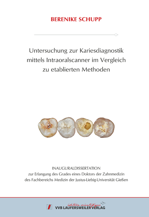
Untersuchung zur Kariesdiagnostik Mittels Intraoralscanner im Vergleich zu Etablierten Methoden
Seiten
2022
VVB Laufersweiler Verlag
978-3-8359-7051-9 (ISBN)
VVB Laufersweiler Verlag
978-3-8359-7051-9 (ISBN)
- Keine Verlagsinformationen verfügbar
- Artikel merken
Das Ziel der vorliegenden Untersuchung war es, die Kariesdiagnostikfunktion dreier Intraoralscanner (IOS) unterschiedlicher Dentalhersteller zu untersuchen und mit etab-lierten Diagnostikmethoden zu vergleichen.
Zur Durchführung der vorliegenden in-vitro Studie wurden aus insgesamt 64 humanen Zähnen des Milchgebisses (n = 13) und des bleibenden Gebisses (n = 51) Modelle zur okklusalen und zur approximalen Kariesdiagnostik hergestellt. Für die Okklusalkari-esdiagnostik wurde ein sechsflächiges Gitternetz angelegt, wodurch 78 Flächen des Milchgebisses sowie 306 Flächen des bleibenden Gebisses beurteilt werden konnten. Zur standardisierten approximalen Kariesdiagnostik wurde jeder Zahn in eine mesio-approximale und in eine disto-approximale Fläche eingeteilt, sodass 26 Flächen des Milchgebisses und 102 Flächen des bleibenden Gebisses untersucht werden konnten.
Die Untersuchung der Flächen erfolgte mittels klinischer Diagnostik – visuell, radio-logischer Diagnostik – Bissflügelaufnahme, Nahinfrarot-Transillumination – DIAGNO-cam (KaVo, Biberach, Deutschland) sowie der IOS TRIOS 4 (3Shape, Kopenhagen, Dänemark), iTero Element 5D (Align Technology, Or Yehuda, Israel) und Planmeca Emerald S (Planmeca Oy, Helsinki, Finnland). Als Referenzmethode fand das µ-CT (TomoScope XS-FOV; Werth Messtechnik GmbH, Gießen, Deutsch-land) Anwendung.
Hinsichtlich ihrer Eignung an Okklusal- und Approximalflächen sowie an Zähnen des Milch- und des bleibenden Gebisses zeigten die untersuchten IOS Differenzen. Der IOS Planmeca Emerald S wies an Zähnen des bleibenden Gebisses teils bessere Er-gebnisse auf als der jeweilige Goldstandard (Reliabilität, Summe aus Sensitivität und Spezifität sowie der logistischen Regression). Im Milchgebiss zeigte hingegen der IOS TRIOS 4 eine größere Reliabilität sowie eine höhere Summe aus Sensitivität und Spezifität als die anderen IOS, sollte jedoch durch den entsprechenden Goldstandard ergänzt werden.
Schlussfolgernd lässt sich sagen, dass in der vorliegenden Untersuchung keine Me-thode ermittelt werden konnte, die an beiden untersuchten Flächen und Dentitionen als alleinige Kariesdiagnostikmethode geeignet wäre. Insgesamt zeigte sich das Kari-esdiagnostiktool von IOS jedoch als sinnvolle Ergänzungsfunktion, deren Validität durch künstliche Intelligenz weiter optimiert werden sollte. The aim of the present study was to investigate the caries diagnostic function of three intraoral scanners (IOS) from different dental manufacturers and to compare them with established diagnostic methods.
To conduct the present in vitro study, models for occlusal and proximal caries diagnosis were prepared from a total of 64 human teeth of the primary dentition (n = 13) and the permanent dentition (n = 51). For occlusal caries diagnosis, a six-surface grid was created, allowing 78 surfaces of the primary dentition and 306 surfaces of the permanent dentition to be assessed. For standardized proximal caries diagnosis, each tooth was divided into a mesio-approximal and a disto-approximal surface, so that 26 surfaces of the primary dentition and 102 surfaces of the permanent dentition could be examined.
The surfaces were examined using clinical diagnostics – visual, radiological diagnostics – bitewing, near-infrared transillumination – DIAGNOcam (KaVo, Biberach, Germany) and the three IOS TRIOS 4 (3Shape, Copenhagen, Denmark), iTero Element 5D (Align Technology, Or Yehuda, Israel) and Planmeca Emerald S (Planmeca Oy, Helsinki, Finland). The µ-CT (TomoScope XS-FOV; Werth Messtechnik GmbH, Giessen, Germany) was used as the reference method.
With regard to their suitability on occlusal and proximal surfaces as well as on teeth of the primary and permanent dentition, the investigated IOS showed differences. The Planmeca Emerald S IOS showed partly better results on permanent dentition teeth than the respective gold standard (reliability, sum of sensitivity and specificity as well as logistic regression). In the primary dentition, the IOS TRIOS 4 showed greater reliability and a higher sum of sensitivity and specificity than the other IOS. However, its use should still be supplemented by the corresponding gold standard.
In conclusion, no method could be identified in the present study that is suitable as the sole caries diagnostic method on both investigated surfaces and dentitions. Overall, the caries diagnostic tool from IOS proved to be a useful supplementary function whose validity should be optimized by artificial intelligence.
| Erscheinungsdatum | 03.08.2022 |
|---|---|
| Reihe/Serie | Edition Scientifique |
| Verlagsort | Gießen |
| Sprache | deutsch |
| Maße | 148 x 210 mm |
| Gewicht | 200 g |
| Themenwelt | Medizin / Pharmazie ► Zahnmedizin ► Klinik und Praxis |
| Schlagworte | Dentalmedizin • Intraoralscanner • Karies |
| ISBN-10 | 3-8359-7051-8 / 3835970518 |
| ISBN-13 | 978-3-8359-7051-9 / 9783835970519 |
| Zustand | Neuware |
| Haben Sie eine Frage zum Produkt? |
Mehr entdecken
aus dem Bereich
aus dem Bereich
Prüfungswissen Kariologie, Endodontologie und Parodontologie
Buch (2023)
Deutscher Ärzteverlag
CHF 97,95


