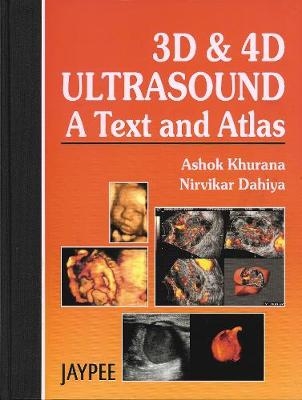
3D and 4D Ultrasound: A Text and Atlas
Seiten
2004
Anshan Ltd (Verlag)
978-1-904798-13-2 (ISBN)
Anshan Ltd (Verlag)
978-1-904798-13-2 (ISBN)
- Titel ist leider vergriffen;
keine Neuauflage - Artikel merken
Three dimensional (3D) and Real Time three dimensional (4D) ultrasound has changed the way scans are carried out. This book deals with Real Time 4D ultrasound, and discusses its usage when applied to all internal organs. It also features over one thousand black and white and colour images.
Three dimensional (3D) and Real Time three dimensional (4D) ultrasound has changed the way scans are carried out. Such ultrasound techniques have now become acceptable as valuable methods of diagnosis. 3D and 4D ultrasound can nowadays be applied to the examination of all internal body organs. Originally developed in the field of obstetrics and gynaecology, ultrasound diagnosis is now common place throughout internal medicine. This book is the first to deal with Real Time 4D ultrasound, and discusses its usage when applied to all internal organs. This book is written by two internationally renowned practitioners of state of the art 3D and 4D ultrasound techniques.
Three dimensional (3D) and Real Time three dimensional (4D) ultrasound has changed the way scans are carried out. Such ultrasound techniques have now become acceptable as valuable methods of diagnosis. 3D and 4D ultrasound can nowadays be applied to the examination of all internal body organs. Originally developed in the field of obstetrics and gynaecology, ultrasound diagnosis is now common place throughout internal medicine. This book is the first to deal with Real Time 4D ultrasound, and discusses its usage when applied to all internal organs. This book is written by two internationally renowned practitioners of state of the art 3D and 4D ultrasound techniques.
Ashok Khurana - Medical Director, The Ultrasound Lab, New Delhi Nirvikar Dahiya - Head of Dept of Ultrasound at KG Hospital, Coimbatore, India
The Physical Principles and Techniques; The Liver and The Biliary Tract; The Pancreas, Para-aortic areas and the Spleen; The Kidneys, Ureters and Bladder, The Prostate; The Small Parts - Breast, Thyroid, Scrotum, and Musculoskeletal Structures; The Vascular Systems; The Endometrium; The Myometrium; The Adnexa; Cancer of the Cervix; Congenital Malformations of the Uterus; Early Pregnancy; Fetal Malformations; The Placenta, Umbilical Cord, Amniotic Fluid and Cervix.
| Erscheint lt. Verlag | 1.8.2004 |
|---|---|
| Zusatzinfo | 1000 Halftones, black and white |
| Verlagsort | Tunbridge Wells |
| Sprache | englisch |
| Maße | 224 x 286 mm |
| Gewicht | 1320 g |
| Themenwelt | Medizinische Fachgebiete ► Radiologie / Bildgebende Verfahren ► Sonographie / Echokardiographie |
| ISBN-10 | 1-904798-13-6 / 1904798136 |
| ISBN-13 | 978-1-904798-13-2 / 9781904798132 |
| Zustand | Neuware |
| Haben Sie eine Frage zum Produkt? |
Mehr entdecken
aus dem Bereich
aus dem Bereich
Begleitbuch für Sonografiekurse, Klinik und Praxis
Buch | Softcover (2023)
Urban & Fischer in Elsevier (Verlag)
CHF 37,80
Organbezogene Darstellung von Grund- und Aufbaukurs sowie …
Buch | Hardcover (2020)
Deutscher Ärzteverlag
CHF 147,30


